ECR 2019 / C-1661
New images in the evaluation of the inferior vena cava: state of the art
Congress:
ECR 2019
Poster Number:
C-1661
Type:
Educational Exhibit
Keywords:
Veins / Vena cava, Vascular, Abdomen, MR-Angiography, CT-Angiography, Image manipulation / Reconstruction, Computer Applications-3D, Education and training
Authors:
M. F. P. Pereira1, E. C. Raimundo1, L. D. P. G. D. Farias2, D. C. Menezes1, I. S. Faé1, M. D. O. M. Hans1, M. V. Galon1, A. F. Pedri1, M. D. S. Guedes1; 1São Paulo/BR, 2São Paulo, SP/BR
DOI:
10.26044/ecr2019/C-1661

Fig. 1:
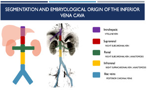
EMBRYOLOGICAL ORIGIN OF THE INFERIOR VENA CAVA

Fig. 2:
SEGMENTATION AND EMBRYOLOGICAL ORIGIN OF THE INFERIOR VENA CAVA
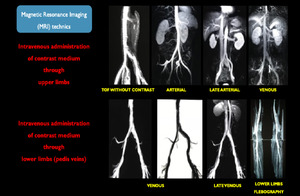
 technics
References: Hospital Alvorada/ Americas Serviços Médicos, São Paulo / Brazil")
Fig. 3:
Magnetic Resonance Imaging (MRI) technics