Intravoxel incoherent motion (IVIM) imaging is a concept used to quantitatively assess all microscopic translational motions from diffusion-weighted magnetic resonance signals.
There are several IVIM models that can estimate the physical properties of biological tissues [1-4].
A typical IVIM model is as follows (also see Figure 4).
a.
Mono-exponential model
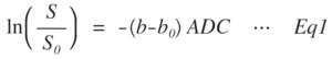
Fig. 1: Mono-exponential model
where the subscript of 0 for b0 and S0 indicates b-value and diffusion-weighted (DW) signal at 0 mm2/s,
respectively.
In addition,
b and S are the desired b-value and DW signal at the same b-value.
ADC means the apparent diffusion coefficients,
and it indicates an amount of diffusion component for each voxel and is calculated from the slope of the natural logarithm of the DW signal attenuation curve at each b-value.
b.
Bi-exponential model
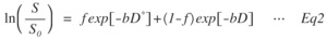
Fig. 2: Bi-exponential model
where f,
D*,
and D are the flowing blood volume fraction,
perfusion-related diffusion,
and diffusion coefficient of water,
respectively.
The bi-exponential model has unique features that can obtain a fast diffusing proton pool and a slow diffusing proton pool for each voxel as D* and D while would lead to estimation errors due to three of free models.
c.
Stretched model
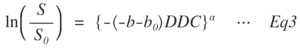
Fig. 3: Stretched-exponential model
DDC denotes mean intravoxel diffusion rates due to the heterogeneity of biological tissue,
and α is the stretch parameter and varies from 0 to 1.
A fitted line of signal attenuation behaves as a mono-exponential model when the value of α is equal to 1,
and the value of α decreases by the addition of multiple separable proton pools within the voxel reflecting the extent of non-Gaussian diffusion [4].
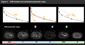
Fig. 4: IVIM models and estimated parameter maps
These IVIM models can obtain unique physical properties and empirically handle non-Gaussian diffusion.
The non-Gaussian diffusion is a diffusion behavior within the voxel and can be observed on the signal attenuation curve of the natural logarithm of the DW signal derived from the b-value [5].
The effect of the non-Gaussian diffusion is expected to be greater at a region of high b-value (200–1500 mm2/s) than that at low b-value (0–200 mm2/s).
It is also expected to provide information about shear stiffness on the liver tissue (see Figure 5),
and is used in virtual magnetic resonance elastography (MRE).
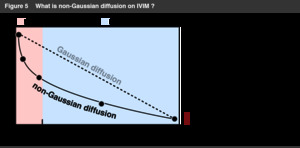
Fig. 5: Non-Gaussian diffusion on IVIM
The virtual MRE procedure was reported by Le Bihan et al.
[6].
It is based on the concept that shear stiffness is related to non-Gaussian diffusion,
and is empirically calculated from the relationship between conventional MRE and the DW signal [6] (Flowchart of calculating virtual shear stiffness is shown in Figure 6).
In a virtual MRE,
an IVIM model that can reliably detect the amount of non-Gaussian diffusion is required to reflect its dependence on the measured changes in shear stiffness.
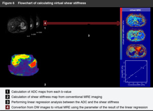
Fig. 6: Flowchart of calculating virtual shear stiffness
In this study,
a relationship between the conventional MRE and the estimated parameter for each IVIM model was investigated to confirm how the non-Gaussian diffusion obtained from each of the IVIM models relates to the shear stiffness on the liver tissue (see Figure 7).
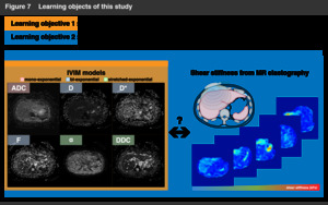
Fig. 7: Learning objects of this study
