ECR 2019 / C-1997
Localization and Lateralization of Memory by BOLD Functional MRI Using Home Town Walking Paradigm for Temporal Lobe Epilepsy Surgery. How I Do It.
Congress:
ECR 2019
Poster Number:
C-1997
Type:
Educational Exhibit
Keywords:
Neuroradiology brain, MR-Functional imaging, Diagnostic procedure, Seizure disorders
Authors:
J. Herbert1, K. Kawsar1, M. Harley1, N. Davies 2, R. Flintham2, A. Zisakis1, D. McCorry1, R. Chelvarajah1, V. Sawlani1; 1Birmingham/UK, 2Birmingham /UK
DOI:
10.26044/ecr2019/C-1997
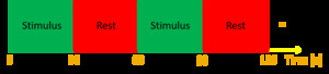
, followed by a period of ‘rest’ in which the haemodynamic response is allowed to return to a resting baseline. Brain activity is averaged across all trials within the block.")
Fig. 1:
Home-town walk memory paradigm - 10 sets of Active-Baseline blocks. A stimulus...

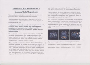
Fig. 2:
Example of the patient information sheet along sent to patients with the...

Fig. 3:
Memory fMRI - Patient Information Sheet Example of Home Town Walking Paradigm

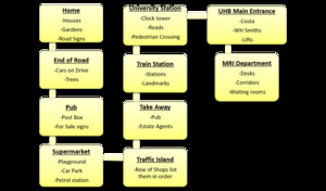
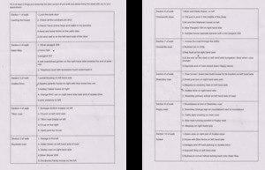
Fig. 4:
Memory fMRI - Completed Patient Crib Sheet - 10 sections

Fig. 5:
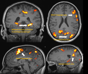
Memory fMRI - Home town walking paradigm showing bilateral activation in the...

Fig. 6:
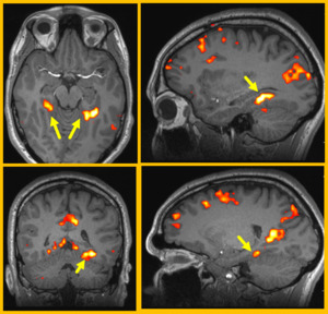
Memory fMRI ‘Home Town Walk’ Paradigm Patient - Day one study shows...

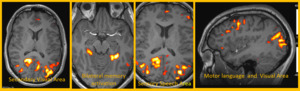
Fig. 7:
Memory fMRI ‘Home Town Walk’ Paradigm Patient - Day two study shows...