ECR 2019 / C-2347

Normal looking abnormal brain: Review areas in routine practice.
Congress:
ECR 2019
Poster Number:
C-2347
Type:
Educational Exhibit
Keywords:
Anatomy, Emergency, Neuroradiology brain, CT, MR, PACS, Audit and standards, Comparative studies, Observer performance, Ischaemia / Infarction, Trauma, Education and training
Authors:
P. Hota, H. C. Chadaga, S. Patwari, S. Kanumukullakshminarayana; Bangalore/IN
DOI:
10.26044/ecr2019/C-2347
. Quality Initiatives: Blind Spots at Brain Imaging. RadioGraphics, 29(7), 1877–1896.")
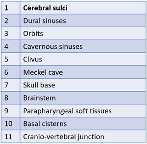
Table 1:
"Blind spots" in Brain imaging

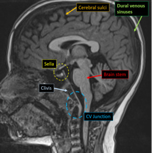
Fig. 41:
Midsagittal section of MRI Brain showing "BLIND SPOTS"
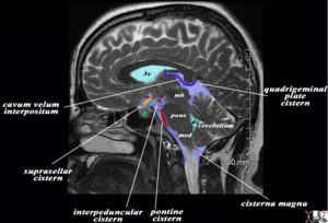
,the ventricles (teal blue), and a variety of cerebral structures including the midbrain (mb) pons, medulla (med) and cerebellum. References: http://www.imagingdomain.com")
Fig. 42:
Sagittal MRI highlighting basal cisterns(light blue),the ventricles (teal...