Type:
Educational Exhibit
Keywords:
Congenital, Intrauterine diagnosis, Ultrasound-Colour Doppler, MR, Thorax, Foetal imaging
Authors:
E. Di Puglia1, T. Fazecas1, R. Nogueira1, H. WERNER JÚNIOR1, P. A. N. Daltro1, B. GUEDES RIBEIRO1, M. G. M. Waksman2, E. Antunes1, C. L. Leidersnaider1; 1Rio de Janeiro/BR, 2Petrópolis, RJ/BR
DOI:
10.26044/ecr2019/C-2640
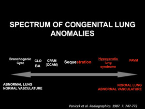
Background
SUMMARY
Congenital lung malformations are rare.
Constitute a heterogeneous group of affections that may involve the lung parenchyma,
bronchi,
and vasculature,
appearing in an isolated or combined form.
They can be detected prenatally,
in infancy or later in childhood,
or even in adult life.
With recent advances in prenatal imaging evaluation,
detection of asymptomatic congenital lung malformations has been increasing in frequency.
It can range from small and asymptomatic entities to large space-occupying masses that require immediate surgical treatment.
ETIOLOGY
It has been suggested that many of pulmonary anomalies present pulmonary dysplastic changes probably due to an obstruction malformation sequence.
Variations in the level,
completeness,
and timing of airway obstruction are suggested as being responsible for the spectrum of anomalies that may happen (figure 2).
The vascular abnormality is also usually suggested as an etiologic or associated feature.
It is important to know that lung congenital lung malformations are commonly interrelated,
and characteristics of different imaging and pathologic entities can coexist in the same lesion or separately in the same child.
Fig. 2: Spectrum of congenital lung anomalies