ECR 2019 / C-2805
PET-CT upstaging of unilateral operable breast cancer and its co-relation with molecular subtypes
Congress:
ECR 2019
Poster Number:
C-2805
Type:
Scientific Exhibit
Keywords:
Breast, Hybrid Imaging, Oncology, PET-CT, Cost-effectiveness, Molecular imaging, Cancer
Authors:
B. Raghavan1, S. Viswanathan1, G. SIVARAMALINGAM2, S. Singh1, R. Baburaj1, B. Ramakrishnan1; 1Chennai/IN, 2CHENNAI, tamilnadu/IN
DOI:
10.26044/ecr2019/C-2805

Fig. 1:
Graphical representation of upstaged and unchanged cases after PET CT.

Fig. 2:
Unilateral operable breast cancer cases upstaged to III C and IV stages by...
 and insignificant axillary lymph nodes-Clinical stage I (T1cN0M0). PET CT showed FDG positive T1c N0 M1 - IV. T1c: Right breast mass in inner lower quadrant with maximum dimension of 1.8 cms (SUVmax:6.6). M1: High metabolic activity in D12 vertebral body with corresponding poorly defined lytic lesion in CT.")
Fig. 3:
45 years old female presented with right breast malignant mass (IHC: ER + PR+...
-Clinical stage IIA(T2N0M0). PET CT showed FDG positive T2N1M1 - IV. T1c: Right breast mass in upper inner quadrant with maximum dimension of 3.9 cms (SUVmax:11.3). N1: prominent axillary nodes. M1:Multiple metastatic cervical, mediastinal and retroperitoneal lymph nodes and skeletal metastases.")
Fig. 4:
44 years old female presented with right breast malignant mass (IHC: ER + PR+...
 -Clinical stage IIA(T2N0M0). PET CT showed FDG positive T2N1M1 - IV. T2: Left breast mass in lower outer quadrant with maximum dimension of 2.8 cms (SUVmax:7.6). N1:Subcentimeter FDG positive left axillary nodes. M1: Hepatic and focal lytic acetabulum and iliac bone metastaes.")
Fig. 5:
Case of 52 years old female with left breast malignant mass ( IHC: ER - PR -...
 and prominent left axillary lymph nodes.
-Clinical stage IIB(T2N1M0). PET CT showed FDG positive T2N1M1-Stage IV. T2: Left breast mass in lower outer quadrant with maximum dimension of 1.6 cms (SUVmax:3.4). N1 : Left axillary lymph nodes. M1: Metastatic mediastinal nodes and bony secondaries.")
Fig. 6:
Case of 63 years old female with left breast cancer( IHC: ER + PR+ Her 2 -...
 and subcentimeter left axillary lymph nodes
-Clinical stage IIB(T2N1M0). PET CT showed FDG positive T2N1M1-Stage IV. T2: Left breast mass in upper central quadran with maximum dimension of 2.1 cms (SUVmax:3.2). N1 :Subcentimeter left axillary lymph nodes.M1: Vermis metastases.")
Fig. 7:
Case of 41 years old female with left breast cancer( IHC: ER - PR+ Her 2 -...

Fig. 8:
Distribution of various metastases newly detected by PET-CT.

Fig. 9:
Sub centimetre right breast primary cancerous nodule. Multiple enlarged...
 PET CT image in a 45 Years old female with a 1.8 cms invasive ductal carcinoma of right breast showing SUVmax:6.6. Immunohistochemistry staining results b)Expression of ER in 95% tumor cells. c) Expression of PR in 95% tumor cells. d) Her 2 neu negative staining with 0 score i.e. absence of membrane staining in tumor cells. e) Low Ki 67 proliferation index: 1-2%.")
Fig. 10:
Luminal A breast cancer: a) PET CT image in a 45 Years old female with a 1.8...
 PET CT image in a 43 Years old female with a 2.2 cms invasive ductal carcinoma of left breast showing SUVmax:5.8. Immunohistochemistry staining results b)Expression of ER in 95% tumor cells. c) Expression of PR in 75% tumor cells. d) Her 2 neu positive staining with 3+ score i.e. complete and strong membrane staining in tumor cells. e) High Ki 67 proliferation index with 70-75% of nuclear staining intumor cells.")
Fig. 11:
Luminal B breast cancer: a) PET CT image in a 43 Years old female with a 2.2...
 PET CT image in a 51 Years old female with a 2.5 cms infiltrating ductal carcinoma of left breast showing moderate FDG uptake(SUVmax:7.6). Immunohistochemistry staining results b)Expression of ER is negative in tumor cells. c) Expression of PR is negative in tumor cells. d) Her 2 neu 3+ score i.e. complete and strong membrane staining in tumor cells.")
Fig. 12:
Her 2 neu positive breast cancer: a) PET CT image in a 51 Years old female with...
 PET CT image in a 52 Years old female with a 2.8 cms invasive ductal carcinoma of left breast showing moderate FDG uptake(SUVmax:7.6). Immunohistochemistry staining results b)Expression of ER is negative in tumor cells. c) Expression of PR is negative in tumor cells. d) Her 2 negative i.e. complete absence of membrane staining in tumor cells.")
Fig. 13:
Triple negative breast cancer: a) PET CT image in a 52 Years old female with a...

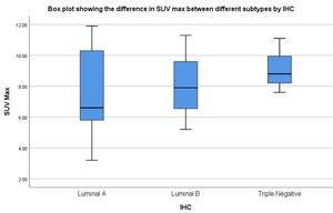
Fig. 14:
Box and whisker plot showing distribution of SUV max in various IHC types