Type:
Educational Exhibit
Keywords:
Neuroradiology spine, MR physics, MR, Education, Physics, Cerebrospinal fluid, Education and training
Authors:
V. B. Pai1, B. S. Purohit1, Y. Y. Sitoh2, K. Gupta3, B. Pai3; 1Singapore /SG, 2SINGAPORE/SG, 3MUMBAI, MA/IN
DOI:
10.26044/ecr2019/C-2890
Background
Cerebrospinal fluid (CSF) is the colourless fluid which encases the neuraxis. It cushions the brain and spinal cord from injury and serves as an important medium for the transport of nutrients and wastes. On an average,
the de novo volume of CSF measures 140 mL.
This is superimposed by an average daily production of 500 mL of CSF produced by specialized ependymal tissues known as the choroid plexus found within the ventricles (1,2).
CSF is in a dynamic state of motion,
circulated from the lateral and third ventricles into the fourth ventricle across the cerebral aqueduct.
CSF exits the fourth ventricle through the median and lateral apertures into the subarachnoid spaces around the cerebral convexities and the spinal cord Finally,
CSF is absorbed by arachnoid granulations into the dural venous system (1).
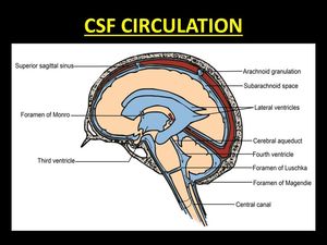
Fig. 1: Diagrammatic representation of the circulation of CSF around the brain and spinal cord.
References: Dr. Vivek Pai
The movement of CSF in the craniospinal spaces is a complex phenomenon.
CSF exits the ventricles in a single outward direction but is multidirectional in the subarachnoid space (3).
CSF circulation is pulsatile,
attributed to the pulsations of the brain caused by the intracranial vessels during the cardiac cycle (1).
It is this physiological movement of CSF which causes artifacts at MR imaging.

