Type:
Educational Exhibit
Keywords:
Tissue characterisation, Pathology, Embolisation, Complications, MR-Diffusion/Perfusion, MR-Angiography, MR, Pelvis, Interventional vascular, Genital / Reproductive system female
Authors:
I. Kavelj, L. Novosel, K. Bolanca, N. BABIC, D. Zadravec; Zagreb/HR
DOI:
10.26044/ecr2019/C-3564
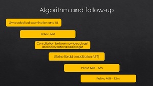
Background
Uterine fibroids (UF),
also referred to as myomas or leiomyomas,
are the most common benign tumors of female pelvis that occur in 20-30% of women in reproductive age.
UF may be single or multiple,
variable in size.
These tumors are composed of smooth muscle cells with variable amounts of fibrous connective tissue.
They are typically asymptomatic but when presented with symptoms (20-50 %) like abnormal uterine bleeding (menometrorrhagia),
pelvic pain or pressure on adjacent organs they may be treated with uterine fibroid embolization (UFE).
After initial examination by gynecologist,
patients with diagnosed leiomyomas are referred to the pelvic MRI,
followed by consultations between the gynecologist and interventional radiologist and possible embolization.
Follow-up MRI is done 6 and 12 months after the procedure (Fig.
1).
Fig. 1: Algorithm of management and follow-up of uterine fibroids in interventional radiology
Among several possible options for treatment of uterine fibroids,
in the last two decades UFE has been recognised as a safe and effective method that reduces or completely eliminates the symptoms associated with these benign tumors,
especially in cases of multiple fibroids and contraindications for surgery.
It is a minimally invasive treatment option that uses microspheres to embolize the branches of uterine arteries that supply uterine fibroids to the visible stasis of contrast flow (Fig.
2).
Fig. 2: 47-years-old female with prolonged and excessive menstrual bleeding underwent uterine fibroid embolization (UFE)
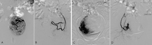
: vascular supply of uterine fibroid by both uterine arteries, dominantly from the left one. Images show embolization of both left (A,B) and right uterine artery (C,D).
Patient selection for UFE is influenced by:
- presenting symptoms,
- physical examination,
- imaging findings and
- patient preferences.