Keywords:
Radiographers, Radioprotection / Radiation dose, Image manipulation / Reconstruction, Education, Technical aspects, Quality assurance
Authors:
C. K. Bandeira1, H. Vieira Neto2, M. P. M. M. Vieira2; 1São José dos Pinhais/BR, 2Curitiba/BR
DOI:
10.26044/ecr2019/C-3675
Methods and materials
The dynamic cardiac phantom was developed by using a cardiac simulator consisting of latex and cannulas that simulate the pulmonary and systemic circulation (Fig.1).
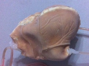
Fig. 1: Cardiac simulator consisting of latex.
References: Physics, Universidade Tecnológica Federal do Paraná - Curitiba/BR
The internal components are showed in the Fig.
2.
The cardiac simulator has four chambers that represent two atria and two ventricles.
A septum separates the chambers between right and left side.
Between the atria and the ventricles are located septa that simulate the atrioventricular valves,
that is,
they allow the passage of fluid from the atria to the ventricles and prevent the circulation in the opposite direction.
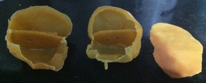
Fig. 2: Internal structures of the cardiac simulator.
References: Physics, Universidade Tecnológica Federal do Paraná - Curitiba/BR
To simulate the small circulation,
a single lumen catheter was connected from the right ventricle to the left atrium and to represent the systemic circulation a double lumen catheter was connected from the left ventricle to the right atrium.
One of these lumens was used to inject fluid into the right atrium.
Four hobby servomotors (Fig.
3) were positioned next to each of the four cardiac chambers of the cardiac phantom in such a way that the horns attached to each servomotor axis compress the respective chambers.
The servomotors were controlled by PWM signals generated by an Arduino Uno.
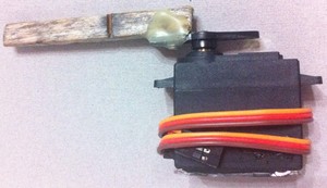
Fig. 3: Hobby servomotor with horns attached to axis used to compress the phantom chambers.
References: Physics, Universidade Tecnológica Federal do Paraná - Curitiba/BR
The amplitude and frequency of rotation of the axis of each servomotor was controlled from a code developed in the Arduino Integrated Development Environment.
For each heartbeat the total rotation range is 90 degrees,
being 45 degrees clockwise and 45 degrees counterclockwise,
and the time for 1 degree rotation is 10 milliseconds.
Movement occurs synchronously.
