Keywords:
Radiographers, Radioprotection / Radiation dose, Image manipulation / Reconstruction, Education, Technical aspects, Quality assurance
Authors:
C. K. Bandeira1, H. Vieira Neto2, M. P. M. M. Vieira2; 1São José dos Pinhais/BR, 2Curitiba/BR
DOI:
10.26044/ecr2019/C-3675
Results
The phantom developed in this work (Fig.
4) resembles dynamic phantoms presented in the literature [2,3] that reproduce the heart rate during the simulation of radiological procedures.
A differential of the presented simulator is the possibility of changing the rotation time of the servomotor horns in the developed code,
which allows simulating pathologies such as cardiac arrhythmias.
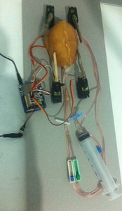
Fig. 4: Developed cardiac phantom. The four hobby servomotors next to each chamber and the syringe used to inject fluid test and iodine-based contrast.
References: Physics, Universidade Tecnológica Federal do Paraná - Curitiba/BR
A radiographic image (Fig.
5) shows the internal components of the phantom.
The image showed in the Fig.
6 was recorded in a fluoroscopy equipment and indicated that injected iodine-based contrast filled the cardiac chambers.
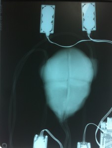
Fig. 5: Radiographic image showing the internal components of the cardiac simulator and the catheters that connect the chambers.
References: Physics, Universidade Tecnológica Federal do Paraná - Curitiba/BR
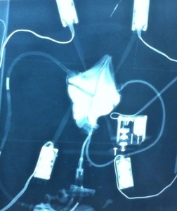
Fig. 6: Fluoroscopy image showing the cardiac chambers filled with iodine-based contrast.
References: Physics, Universidade Tecnológica Federal do Paraná - Curitiba/BR
The Arduino code developed allowed controlling the movement of the horns of each servomotor independently.
The compression of the atriums and ventricles occurred at a frequency of 1 hertz.
However,
the intensity of compression of the cardiac chambers was not enough to completely drain them out,
which resulted in low flow velocity.
There was also a small fluid return through the cannulas that connect the ventricles to the atrium.
