Learning objectives
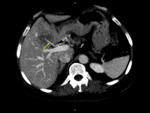
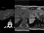
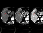
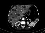
To study the etiology, pathophysiology and to describe US, CT and MRI appearance of hepatic pseudolesions.
To recognize vascular anatomical variants on various imaging studies.
To discriminate pseudolesions from true lesions.
Background
Pseudolesions of the liver are non-neoplastic abnormalities and can be divided into parenchymal and vascular changes. Pesudolesions often occur due to blood flow abnormalities and can be found in both cirrhotic and non-cirrhotic livers.[1]
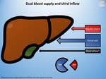
The liver has dual blood supply with the portal vein carrying 75% of blood and the hepatic artery with 25% of blood and a drainage route through the hepatic veins. Both systems have communications between them that allow supporting mechanisms.[1]
There are anatomical variations of liver blood vessels of which only...
Findings and procedure details
TABLE OF CONTENTS
-Parenchymal Pseudolesions
oFocal Fatty Liver and Focal Spared Areas
oInflammatory Pseudotumors (IPT)
oPeliosis Hepatis
oConfluent Hepatic Fibrosis
oSegmental Hypertrophy
oParenchymal Compression
-Vascular Pseudolesions
oTransient Hepatic Attenuation/Intensity Differences (THAD/THID)
oVascular Malformations
Parenchymal Pseudolesions
Focal Fatty Liver and Focal Spared Areas
Fat accumulation is one of the most common conditions of the liver. Fatty liver is characterized by the accumulation of triglyceride droplets in hepatocytes not in the extracellular matrix.[4]
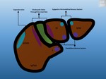
Focal Fatty Change (FFC) is related to veins of third inflow(Fig. 3). These veins...
Conclusion
In patients with cancer, some forms of pseudolesions can cause panic, therefore, in-depth knowledge of liver hemodynamics and enhancement patterns is important in judging whether an abnormal finding is induced by a true lesion.
Although pseudolesions tend to have a typical appearance, there are many cases with atypical patterns. CT and ultrasonography are useful for the diagnosis of pseudolesions but MRI appears to be
the more sensitive in solving diagnostic problems.
Personal information and conflict of interest
A. V. Neagu; Bucharest/RO - nothing to disclose C.-I. Betianu; Bucharest/RO - nothing to disclose
References
1.Schneider G, Grazioli SL, Saini S, eds. Hepatic pseudolesions. In: MRI of the Liver: Imaging Techniques, Contrast Enhancement, Differential Diagnosis. 2nd edn. Milan: Springer-Verlag Italia 2006: 152-185.
2.Kobayashi, Satoshi & Gabata, Toshifumi & Matsui, Osamu. (2010). Radiologic manifestation of hepatic pseudolesions and pseudotumors in the third inflow area. Imaging in Medicine. 2. 10.2217/iim.10.50.
3.Itai Y, Matsui O.'Nonportal' splanchnic venous supply to the liver: abnormal findings on CT, US and MRI.Eur Radiol1999;9:237-43. 10.1007/s003300050661.
4.Hamer OW, Aguirre DA, Casola G, Lavine JE, Woenckhaus M, Sirlin CB (2006)...


