Actinomycosis usually presents as [4,5]:
- an inflammatory mass;
- abscess, fistula, draining sinus;
- dense fibrosis.
Contrast-enhanced computed tomography allows the assessment of the anatomical location and disease extension, being also useful in monitoring the response to therapy.
The most distinctive CT feature of actinomycosis is the aggressive infiltrative pattern across normal fascial and connective tissue planes - as a result of proteolytic enzymes produced by Actinomyces [5]. Thus, actinomycosis represents one of the most misdiagnosed diseases. Its presentation is more often associated with malignancy, rather than with an infectious process, patients being in most cases referred to surgery [6].
The CT characteristics of abdominopelvic actinomycosis are variable depending on the involvement sites.
Gastrointestinal involvement
CT imaging features:
- concentric bowel wall-thickening with homogenous enhancement and adjacent marked inflammatory fat stranding;
- adjacent solid or cystic mass with irregular margins; solid components of the mass and the surrounding soft-tissue strands tend to show strong enhancement after contrast material administration due to the dense fibrous content – a hallmark of actinomycosis [4,7]. Through contiguous spread, actinomycosis can extend into the omentum and abdominal wall (Fig. 2).
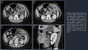
Fig. 2: Case 1. Abdominal actinomycosis of the transverse colon with invasion of the omentum, anterior abdominal wall and gastric wall – mimicking malignancy.
References: Regional Center of Gastroenterology and Hepatology, Department of Radiology - Cluj- Napoca/RO
Other possible complications are fistula and abscess formation with extension into the sacral canal, especially in cases that involve the rectosigmoid colon (Fig. 3).
Differential diagnosis:
- Malignant tumor with necrosis
- Inflammatory bowel disease (Crohn’s disease, ulcerative colitis)
- Intestinal tuberculosis
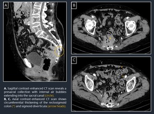
Fig. 3: Case 2. Pelvic actinomycosis of the rectosigmoid colon, complicated with presacral abscess extended into the sacral canal.
References: Regional Center of Gastroenterology and Hepatology, Department of Radiology - Cluj- Napoca/RO
Liver involvement
The liver is rarely involved in abdominal actinomycosis (around 15% of the cases) and it is most commonly secondarily affected as a result of dissemination from other sites [8].
CT imaging features:
Hepatic actinomycosis can present either as a solitary mass-like lesion (usually of low density, with ill-defined margin and hypoenhancement – Fig. 4, Fig. 5) or as single/multiple abscesses with no particular radiographic features compared to liver abscesses of other causes [8,9].
Differential diagnosis:
- Primary hepatic tumor
- Metastatic lesion
- Acute bacterial liver abscess
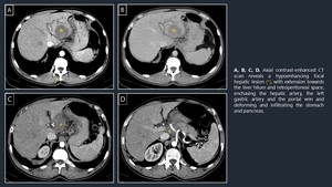
Fig. 4: Case 3. Pseudotumoral aspect of hepatic actinomycosis secondary to gastric perforation caused by the accidental ingestion of a foreign body
References: Regional Center of Gastroenterology and Hepatology, Department of Radiology - Cluj- Napoca/RO
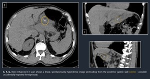
Fig. 5: Case 3. Pseudotumoral aspect of hepatic actinomycosis secondary to gastric perforation caused by the accidental ingestion of a foreign body
References: Regional Center of Gastroenterology and Hepatology, Department of Radiology - Cluj- Napoca/RO
Pelvic organ involvement
Most frequently involved pelvic organs are the ovary and fallopian tube [4]. The long-term use of an intrauterine contraceptive device (IUD) increases the risk of developing pelvic actinomycosis [10].
CT imaging features [4,11,12]:
- adnexal pseudotumoral masses containing suppurative necrotic areas, as well as solid areas strongly enhancing following contrast media administration; adjacent marked inflammatory fat stranding;
- abscesses originating from the ovary, with rim enhancement (Fig. 6); associated extensive fibrosis that could lead to the formation of dense adhesions between small bowel loops;
- due to its infiltrative pattern, pelvic actinomycosis can extensively spread to the adjacent organs leading to the “frozen pelvis” aspect, which favors malignancy rather than an inflammatory process;
- hydronephrosis can be present if the urinary tract is involved.
Differential diagnosis:
- Malignant ovarian tumors
- Endometriosis
- Colonic cancer
- Uterine cancer
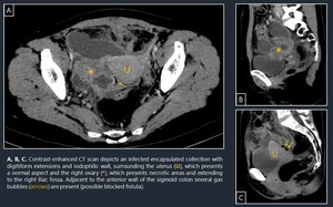
Fig. 6: Case 4. Pelvic actinomycosis presenting as an abscess of the right ovary and Fallopian tube with extension to the last ileal loop and sigmoid colon.
References: Regional Center of Gastroenterology and Hepatology, Department of Radiology - Cluj- Napoca/RO
Key points in differentiating abdominopelvic actinomycosis from malignancy [4]:
- Regional lymphadenopathy is less commonly present in actinomycosis and ascites is either absent or in reduced amounts.
- Actinomycosis is less likely to affect the entire peritoneal cavity, despite its infiltrative pattern.


