Purpose
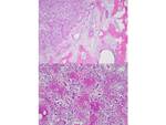
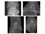
Osteoid osteoma (OO) is a benign bone producing tumor, first described by Jaffe in 1953 [1]. It accounts for 2– 3% of all bone tumors and 10–20% of benign bone tumors and is characterized radiologically by an intracortical nidus, with a variable amount of calcification, as well as cortical thickening, sclerosis, and bone marrow edema [2,3]. Fig. 1The nidus has self-limiting growth, usually becomes asymptomatic and spontaneously heals [4,5].Fig. 2
The typical clinical presentation of OO consists of pain that worsens at night and is...
Methods and materials
From January 2005 to September 2019, 117 patients with OO were studied retrospectively. Patients were selected from a prospectively maintained tumor database using either histological (for those patients undergoing surgery) or radiological diagnosis of OO. Imaging examinations performed during this period were analyzed for the presence of typical imaging findings (CT and MRI) of OO.
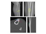
The study included 39 females and 78 males with the mean patient age 22.5 years (age range 2 to 60 years). Fig. 7
Results
CT was performed in 97 patients, MRI in 106 patients. Table 1
The lesions were most frequently located in the tibia (n=35; 30.4%), the femur (n=34; 29.6%), the humerus (n=9; 7.8%), and the radius (n=5; 4.3%). The rest of the lesions (n=44, 37.6%) were located in hands, sacrum, spine, feet, scapula, fibula, patella, ulna and acetabulum. Lesions were located cortically in 81% of the cases. In 1 patient the lesions were multifocal.Table 2 Table 3The mean diameter of the nidus was 9.1 ± 5.4 mm...
Conclusion
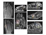
CT is superior to MRI in demonstrating the nidus in OO. It may be usefulfor the identification of the tumor origin and the assessment of the relationship of the tumor to the medulla. In selected cases, in juxta-articular located tumors, reactive bone changes are lacking, and predominantly synovitis may be present – a feature that can be appreciated using MRI [11,15-17]. Due to a variety of radio-morphological presentations, in the majority of cases, the combination of CT and MRI can establish the diagnosis of OO...
Personal information and conflict of interest
J. Igrec; Graz/AT - nothing to disclose I. Brcic; Graz/AT - nothing to disclose M. Bergovec; Graz/AT - nothing to disclose R. Igrec; Graz/AT - nothing to disclose M. A. Smolle; Graz/AT - nothing to disclose B. Liegl-Atzwanger; Graz/AT - nothing to disclose M. Fuchsjäger; Graz/AT - nothing to disclose A. Leithner; Graz/AT - nothing to disclose H. R. Portugaller; Graz/AT - nothing to disclose
References
1.Jaffe HL. Osteoid-osteoma. Proc R Soc Med 1953; 46(12):1007–1012.
2.Chai JW, Hong SH, Choi J-Y, Koh YH, Lee JW, Choi J-A, et al. Radiologic diagnosis of osteoid osteoma: from simple to challenging findings. RadioGraphics. 2010;30(3):737-49.
3.Ghanem I. The management of osteoid osteoma: updates and controversies. Current opinion in pediatrics. 2006;18(1):36-41.
4.Noordin S, Allana S, Hilal K, et al. Osteoid osteoma: Contemporary management.Orthop Rev (Pavia). 2018;10(3):7496. Published 2018 Sep 25. doi:10.4081/or.2018.7496
5.Jo VY, Fletcher CD. WHO classification of soft tissue tumours: an update based on the...


