Purpose
The purpose of the study was to evaluate the hippocampal volume and Alzheimer's disease cortical signature region quantitative measurements in participants with normal cognition (NC), mild cognitive impairment (MCI), and dementia (D).
Methods and materials
In total 44 Participants with suspected cognitive impairment were admitted to the neurologist and the Montreal Cognitive Assessment (MoCA)5 was carried out. Participants were divided into 3 groups:
11 participants with normal cognition (MoCA scores≥26; Average MoCA scores 27.3, SD 1.0; average age 73.2, SD 5.5),
23 participants with mild cognitive impairment (MoCA scores≥18 and ≤25; Average MoCA scores 22.4, SD 2.4; average age 72.3, SD 6.5),
10 participants with dementia (MoCA scores≤17; Average MoCA scores 10.1, SD 4.1; average age 74.8, SD 10.4).
MRI...
Results
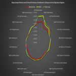
To make an initial evaluation of obtained hippocampal volume values and Alzheimer's disease cortical signature values we made a spider diagram, that could be seen in Figure 2. [Fig 2]
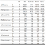
By performing the Kruskal-Wallis test we found statistically significant (p<0.05) differences between groups in:
left hippocampus and right hippocampus where statistically significant differences were observed between D-MCI and D-NC groups, but no statistically significant differences between MCI-NC groups,
left entorhinal cortexand right entorhinal cortex where statistically significant differences were observed between D-MCI and D-NC groups,...
Conclusion
Additional quantitative measures that could be retained from brain MRI scans could be used as additional biomarkers for cognitive impairment diagnostics. Thus facilitating multiparametric brain MRI evaluation in patients with cognitive impairment, including not only qualitative evaluation but also quantitative measurements.
Personal information and conflict of interest
N. Zdanovskis:
Nothing to disclose
A. Platkajis:
Nothing to disclose
A. Kostiks:
Nothing to disclose
K. Šneidere:
Nothing to disclose
A. Stepens:
Nothing to disclose
G. Karelis:
Nothing to disclose
References
1. Dickerson BC, Bakkour A, Salat DH, et al. The cortical signature of Alzheimer's disease: regionally specific cortical thinning relates to symptom severity in very mild to mild AD dementia and is detectable in asymptomatic amyloid-positive individuals.Cereb Cortex. 2009;19(3):497-510. doi:10.1093/cercor/bhn113
2. Busovaca E, Zimmerman ME, Meier IB, et al. Is the Alzheimer's disease cortical thickness signature a biological marker for memory?.Brain Imaging Behav. 2016;10(2):517-523. doi:10.1007/s11682-015-9413-5
3. Dale AM, Fischl B, Sereno MI. Cortical surface-based analysis. I. Segmentation and surface reconstruction. Neuroimage. 1999 Feb;9(2):179-94. doi: 10.1006/nimg.1998.0395....


