Keywords:
Neuroradiology brain, MR, MR-Diffusion/Perfusion, Computer Applications-3D, Computer Applications-Detection, diagnosis, Computer Applications-General, Dementia
Authors:
N. Zdanovskis, A. Platkajis, A. Kostiks, K. Šneidere, A. Stepens, G. Karelis
DOI:
10.26044/ecr2022/C-10232
Methods and materials
In total 44 Participants with suspected cognitive impairment were admitted to the neurologist and the Montreal Cognitive Assessment (MoCA)5 was carried out. Participants were divided into 3 groups:
- 11 participants with normal cognition (MoCA scores≥26; Average MoCA scores 27.3, SD 1.0; average age 73.2, SD 5.5),
- 23 participants with mild cognitive impairment (MoCA scores≥18 and ≤25; Average MoCA scores 22.4, SD 2.4; average age 72.3, SD 6.5),
- 10 participants with dementia (MoCA scores≤17; Average MoCA scores 10.1, SD 4.1; average age 74.8, SD 10.4).
MRI scans were performed on a 3T scanner. Post-processing with cortical reconstruction and volumetric segmentation was performed with the Freesurfer image analysis suite, which is documented and freely available for download online (http://surfer.nmr.mgh.harvard.edu/).3, 4
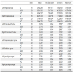
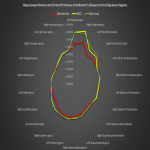
We evaluated hippocampal volume and Alzheimer's disease cortical signature regions and performed the Kruskal-Wallis test with post hoc analysis. Alzheimer's disease cortical signature regions1,2 included:
- the entorhinal cortex,
- temporal lobe medial aspect (separately analyzing fusiform gyrus and parahippocampal gyrus),
- inferior temporal gyrus,
- superior frontal gyrus,
- inferior frontal gyrus (separately analyzing orbital, triangular, and opercular gyrus),
- supramarginal gyrus,
- superior parietal lobule,
- precuneus.


