Purpose
Deep vein thrombosis (DVT) is a blood clot of the deep veins, most commonly of the lower limbs, which can potentially lead to a deadly pulmonary embolism (PE). The diagnosis of a DVT is made in a multi-step process consisting of clinical scores, blood-tests, and the gold-standard compression ultrasound examination of the leg veins. This exam is usually performed by a highly trained radiologist or sonographer [1, 2]. Patients often occupy valuable resources and beds in emergency departments while waiting for a scan or must...
Methods and materials
Patients with a suspected DVT were recruited at a German tertiary care clinic via the emergency department or primary care referral. Each patient underwent two DVT examinations. The first was performed by one of two medical technicians without formal DVT-ultrasound experience or training. Subsequently each patient underwent a full leg ultrasound examination by a highly-trained angiology consultant with 26 years of experience with DVT ultrasound diagnosis.
During the diagnostic scan, the leg was scanned with multiple compressions along the veins from the inguinal crease to...
Results
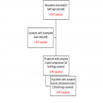
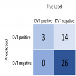
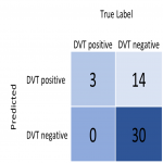
40 patients were included in the study, of which 13 patients received scanning of both legs. 47 complete 2-point DVT-scans from 37 patients, including 43 complete 3-point DVT-scans from 33 patients were collected and uploaded to the platform. 4 patients showed positive results for DVT in the gold standard, on-site diagnostic scan (7.1%) of which full scans were acquired for 3 patients (see figure 8).
[Fig 8]
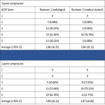
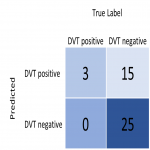
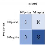
For 3-point compression scans, both raters showed a 100% sensitivity for DVT-diagnosis, with specificities of 62.50% and 65.00%...
Conclusion
Machine learning methods can safely aid non-experts in acquiring valid ultrasound images of venous compressions. These can then be externally reviewed for final expert triage and potential diagnosis, resulting in a very high NPV. This ensures that all patients receiving a negative result from the remote expert were in fact DVT-negative. Over 90% of scans taken by a non-expert with the help of AutoDVT were of diagnostic image quality, this increased to 100% with 2-point compression exams excluding the thigh. This study constituted a small...
Personal information and conflict of interest
J. Oppenheimer:
Employee: ThinkSono GmbH
R. Mandegaran:
Consultant: ThinkSono GmbH
B. Kainz:
Consultant: ThinkSono GmbH
M. P. Heinrich:
Consultant: ThinkSono GmbH
F. Noor:
CEO: ThinkSono GmbH
S. Mischkewitz:
Founder: ThinkSono GmbH
A. Ruttloff:
Nothing to disclose
P. Klein-Weigel:
Nothing to disclose
References
Wells, P.S., et al., Evaluation of D-dimer in the diagnosis of suspected deep-vein thrombosis. N Engl J Med, 2003. 349(13): p. 1227-35.
Lensing, A.W., et al., Detection of deep-vein thrombosis by real-time B-mode ultrasonography. N Engl J Med, 1989. 320(6): p. 342-5.
Needlemann, L., et al., Ultrasound for Lower Extremity Deep Venous Thrombosis: Multidisciplinary Recommendations From the Society of Radiologists in Ultrasound Consensus Conference. Circulation, 2018 137(14): p. 1505-1515.
Liu, R.B., et al., Emergency Ultrasound Standard Reporting Guidelines, American College of Emergency Physicians, 2018: acep.org....


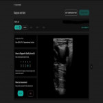

![Fig 7: Table 1: Definition of ACEP-Scores, from: [3]](https://epos.myesr.org/posterimage/esr/ecr2022/159525/media/908715?maxheight=150&maxwidth=150)
