Purpose
To perform a descriptive retrospective study in a tertiary care hospital of patients diagnosed with congenital adrenal lesion in a 10-year period.
To identify suprarenal masses in fetal and neonatal ultrasound (US) and Magnetic Resonance (MR) to orientate their characterization and diagnosis.
Methods and materials
Congenital lesions in suprarenal region can arise from different organs and can be benign or malignant. Fetal and neonatal US and MR can detect suprarenal masses and orientate their characterization and diagnosis.
We performed a descriptive retrospective study in a tertiary care hospital of 55 patients diagnosed with congenital mass in suprarenal area antenatally (beyond 20 weeks of pregnancy) until the first 90 days after birth, from 2011 to 2021. Every patient had a fetal US at week 20 of gestation, and posteriorly US at...
Results
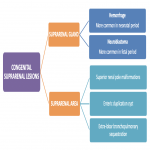
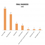
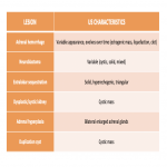
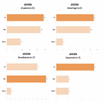
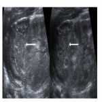
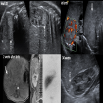
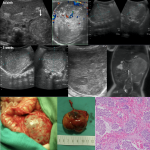
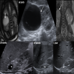
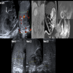
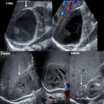
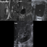
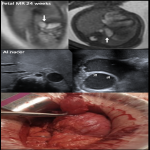
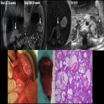
Congenital masses in suprarenal area can be lesions that arise from adrenal gland and lesions from neighboring organs [1].[Fig 1] We retrospectively reviewed 55 patients with congenital suprarenal. 31 were girls and 24 were boys. Most frequent final diagnosis was hemorrhage (n=23), followed by neuroblastoma (n=17).[Fig 2]
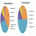
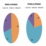
27 cases were diagnosed antenatally on ultrasound study between week 20 and week 40, while 28 cases were identified after birth on postnatal ultrasound studies between day 0 and 90 days after birth. Fetal MR complemented US in...
Conclusion
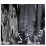
The adrenal gland can be visualized at obstetric US beyond 13 weeks of gestation, being large and globular. In neonates, adrenal gland on US shows differentiation between cortical and medulla, smooth undulations and characteristic shape of two legs [2]. [Fig 9] With child growing, corticomedullary differentiation diminishes by 6 months of age. Beyond this age, the visualization of adrenal glands on US may be difficult. Recognizing the normal shape of adrenal glands in neonates is important because sometimes a lesion does not appear as focal,...
Personal information and conflict of interest
C. C. Sangüesa Nebot:
Nothing to disclose
D. Veiga Canuto:
Nothing to disclose
R. Llorens-Salvador:
Nothing to disclose
References
Maki E, Oh K, Rogers S, Sohaey R. Imaging and Differential Diagnosis of Suprarenal Masses in the Fetus. J Ultrasound Med. mayo de 2014;33(5):895-904.
Sargar KM, Khanna G, Hulett Bowling R. Imaging of Nonmalignant Adrenal Lesions in Children. RadioGraphics. octubre de 2017;37(6):1648-64.
Flanagan SM, Rubesova E, Jaramillo D, Barth RA. Fetal suprarenal masses – assessing the complementary role of magnetic resonance and ultrasound for diagnosis. Pediatr Radiol. febrero de 2016;46[2]:246-54.
Balassy C, Navarro OM, Daneman A. Adrenal Masses in Children. Radiol Clin North Am. julio...


