Keywords:
Foetal imaging, Paediatric, MR, Ultrasound, Education, eLearning, Congenital, Foetus, Haemorrhage
Authors:
C. C. Sangüesa Nebot, D. Veiga Canuto, R. Llorens-Salvador
DOI:
10.26044/ecr2022/C-13280
Methods and materials
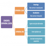
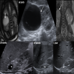
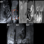
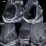
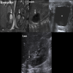
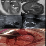
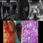
Congenital lesions in suprarenal region can arise from different organs and can be benign or malignant. Fetal and neonatal US and MR can detect suprarenal masses and orientate their characterization and diagnosis.
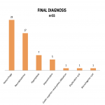
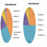
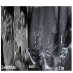
We performed a descriptive retrospective study in a tertiary care hospital of 55 patients diagnosed with congenital mass in suprarenal area antenatally (beyond 20 weeks of pregnancy) until the first 90 days after birth, from 2011 to 2021. Every patient had a fetal US at week 20 of gestation, and posteriorly US at birth depending on initial findings. Fetal MR was performed in cases that required it.
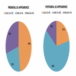
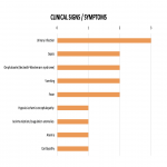
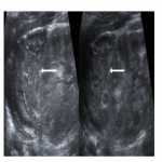
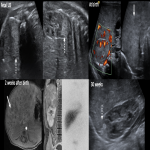
We evaluated clinical histories and imaging studies and collected descriptive variables: age at diagnosis, sex. From prenatal and postnatal US studies we reviewed location (side), size (<5cm/ =or> than 5cm), ultrasonographic pattern (cystic/solid/mixed). On postnatal studies we evaluated Doppler vascularization and suspected diagnosis, and also reviewed newborns clinical signs and symptoms.
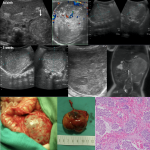
Final diagnosis was confirmed by histological analysis (if surgery/biopsy was performed), or achieved on US follow-up.


