Learning objectives
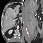
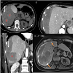
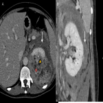
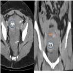
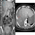
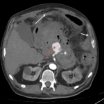
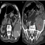
Review the most common causes of hemoperitoneum and illustrate the main imaging findings.
Background
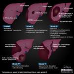
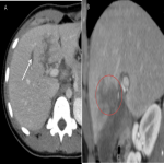
Hemoperitoneum consists of intra-abdominal bleeding and is usually associated with urgent conditions. The main bleeding sources can be divided into two main groups: traumatic with solid organ injury and nontraumatic. In the nontraumatic group, the main etiologies can be divided into tumor-associated hemorrhage, gynecologic conditions, vascular lesions and iatrogenic injuries.
Due to the rapid acquisition time and wide availability, Computed Tomography (CT) assumes a key role in identifying the cause and extent of the hemorrhage.
Findings and procedure details
Hemoperitoneum on CT - main findings and signs
Fluid density and configuration
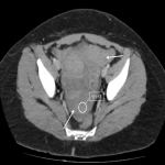
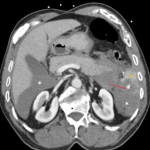
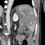
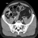
One can distinguish free intraperitoneal fluid from hemoperitoneum by measuring the fluid density. On unenhanced images, unclotted blood is hyperdense (around 30-45 HU), compared to simple free fluid (< 15 HU), due to its high protein content. In the first few hours of hemorrhage, as blood begins to clot, the density increases (HU > 60), with geographic areas of high density surrounded by areas of low density. [1,2]
[Fig 1]
With time and...
Conclusion
It is important that the radiologist becomes aware of the different etiologies of hemoperitoneum and their main CT findings in order to make a timely diagnosis.
Personal information and conflict of interest
F. Grilo:
Nothing to disclose
D. M. R. Barros:
Nothing to disclose
A. C. G. Costa:
Nothing to disclose
F. Vilaverde:
Nothing to disclose
References
[1] Lubner M, Menias C, Rucker C, et al. Blood in the belly: CT findings of hemoperitoneum. Radiographics. 2007;27(1):109-125. doi:10.1148/rg.271065042
[2] Furlan A, Fakhran S, Federle MP. Spontaneous abdominal hemorrhage: causes, CT findings, and clinical implications. AJR Am J Roentgenol. 2009;193(4):1077-1087. doi:10.2214/AJR.08.2231


