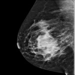
This study population consisted mostly of women with non - palpable lesions (70%). Palpable on physical exam lesions were not associated with microcalcifications detected by mammography (p=0.2458). Notably, calcifications in ductal carcinoma in situ were observed in 80-90% of analyzed lesions on mammography; and predominantly detected in younger women with higher tumor grade. Malignant lesions were associated with irregular, pleomorphic type calcifications (p=0.0084) (Figure 1).
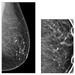
The higher grade of cancer determined by histopathology was strongly associated (p= 0.0401) with grouped or clustered; and segmental distribution of microcalcifications, which was presenting immediate concern as this suggested deposits of calcium within a duct and its branches, potentially caused by multifocal cancer within that breast segment as shown on Figure 2.
Calcification of fibroadenoma were detected less frequently (16%). Notably, in postmenopausal group fibroadenomas regressed, hyalinised with coarse calcifications observed (not shown).
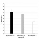
Elevated T1/T2 means of 7.14 ± 1.05 (N=12) were observed for malignant lesions and 6.49 ± 0.63 (N=33) for lesions that were treated prior to qMRI with chemotherapy and/or radiation, as compared to 3.97 ± 0.43 (N=39) for benign lesions (Figure 3).
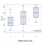
Also, higher means of Wilcoxon score for T1/T2 ratios obtained by qMRI were associated with suspicious for malignancy microcalcifications with worrisome segmental, grouped/ clustered or branching in the duct patterns detected by mammography. This correlation was statistically significant (p=0.0135), as shown on Figure 4.
The higher grade of cancer determined by histopathology was strongly associated (p= 0.0401) with grouped or clustered; and segmental distribution of microcalcifications.
The higher stage of cancer determined by histopathology was strongly associated with clustered, pleomorphic microcalcifications and elevated T1/T2 ratios on qMRI (p= 0.0198). Estrogen, progesterone and Her2/neu triple negative receptors status was strongly correlated with higher T1/T2 ratio (p=0.0019, p=0.0021, and p=0.0030; respectively for each receptor).
Association of palpable mass with calcification was not statistically significant (p=0.245), meaning that routine screening by mammography and/or MRI is still essential to detecting and evaluating suspicious, non-palpable lesions.


