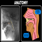
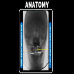
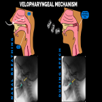
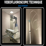
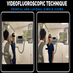
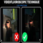
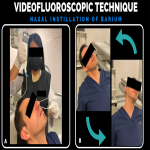
Normal velopharyngeal function depends on three major components which are: anatomy, physiology, and speech learning. Velopharyngeal closure requires normal velopharyngeal structure and adequate velopharyngeal function (neurophysiology), however, is often forgotten that the velopharyngeal valve is an articulator, just like the tongue and lips. As such, there is also a learned component to velopharyngeal function [1].
Velopharyngeal dysfunction (VPD) is known as the condition where the velopharyngeal port does not close completely during the production of oral sounds is used as a broad term that includes all disorders (with various causes) that affect the closure of the velopharyngeal spchinter[1].
Velopharyngeal insufficiency (VPI): Can be defined as an anatomic or structural defect that leads to incomplete closure of the velopharyngeal valve.
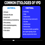
The patient can have complications in eating and drinking, stigmatizing speech with compensatory grimacing, diminished performance in school; and decreased quality of life [2], etiology ranges from abnormalities of the lymphoid tissue to congenital anomalies like such as Treacher-Collins, DiGeorge, and Pierre- Robin syndromes and acquired defects following various surgical procedures, see (Table 1).
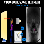
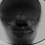
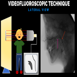
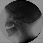
Complete evaluation in these patients includes clinical, radiological, speech analysis, and videonasopharyngoscopy.Among the several imaging methods, videofluoroscopy (VF) can be used to evaluate the dynamic motion of the velopharyngeal valve, it has certain advantages in comparison with cine MRI, like the accessibility of a fluoroscopy device, costs, and a good correlation with video nasopharyngeal endoscopy findings. This study brings accurate information regarding velopharyngeal closure and gives size measurements of velopharyngeal structures and their movements during speech
Radiologists and trainees play a crucial role in the characterization of these imaging findings which can be useful for the surgeon when planning corrective surgery. We aim to provide the key point for a better understanding of the velopharyngeal sphincter anatomy, a step-by-step guide on how to perform, videofluoroscopy, and how to elaborate a complete radiological report.
Radiologists and trainees play a crucial role in the characterization of these imaging findings which can be useful for the surgeon when planning corrective surgery. We aim to provide the key points for a better understanding of the velopharyngeal sphincter anatomy, a step-by-step guide on how to perform videofluoroscopy, and how to elaborate a complete radiological report


