1.- ANATOMY AND PHYSIOLOGY
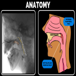
The velopharyngeal sphincter is constituted primarily of a muscular valve that extends from the posterior surface of the hard palate (roof of mouth)to the posterior pharyngeal wall. It is composed of three major elements:
- The velum, also known as soft palate. See (Figure 1).
- Posterior pharyngeal wall (back wall of the throat) [3]. See (Figure 1).
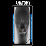
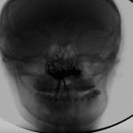
- Lateral pharyngeal walls (sides of the throat). See (Figure 2).
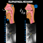
The function of this mechanism is to create a tight seal by elevating the velum and constricting both the lateral and posterior pharyngeal walls to separate the oral and nasal cavities, controlling the airflow to the nose and mouth. See (Figure 3).
Closure of the velopharyngeal sphincter is principally made by retraction and elevation of the soft palate; lateral pharyngeal walls approach each other towards midline and the posterior pharyngeal wall contributes by moving forward to some degree to achieve contact.
Some children make velar contact with the adenoid pad before its involution in adolescence.
Passavant’s ridge is a shelflike musculo-mucosal structure that protrudes forward while the patient is talking and disappears at rest, secondary to contraction of the inferior fibers of the superior constrictor muscle and the horizontal fibers of the palatopharyngeus muscle.
Velopharyngeal muscles include the levator veli palatini, musculus uvulae, tensor veli palatini, superior pharyngeal constrictor, palatopharyngeus, palatoglossus, and salpingopharyngeus that are better assessed by MRI imaging.
2.- VIDEOFLUOROSCOPIC TECHNIQUE.
Videofluoroscopy provides dynamic evaluation by providing actual size measurements of the structures and movements of the velopharyngeal valve while the patient is talking. It also provides dynamic visualization of the entire area of both lateral and posterior pharyngeal walls. Finally, it gives important information about the depth of velopharyngeal closure during speech.[4]
The use of simultaneous audio recording during videofluoroscopic imaging acquisition is essential for a dedicated speech evaluation, but for evaluating velopharyngeal closure pattern, it can be considered optional.
A description on how to perform videofluoroscopy of the velopharyngeal sphincter is listed below.
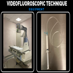
-Equipment: Standard fluoroscopic/radiographic system that is used for the routine examination of the gastrointestinal tract and barium connected to a pediatric feeding tube (See figure 4).
- Patient arrive at the fluoroscopy room, children younger than 10 years must have a companion to be with them during the study, radiological protection must be provided
- The study should be simple enough to be performed on any patient cooperative enough to speak to the examiner. By age 4 most children can cooperate well enough for the radiologist to obtain a good study.
- The procedure must be explained and take a few minutes for the patient to practice a short speech provided preferably by a Speech and Language Pathologist.
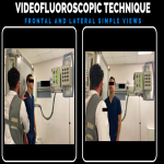
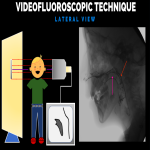
- Frontal and lateral simple views are required. (Other views like oblique, base, and Towne may be added by the radiologist in charge). To display each of these views, the x-ray beam must be oriented perpendicular to the plane of the particular view. See (Figure 5).
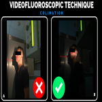
- Beam collimation in all views to limit the X ray beam in the anatomic region of interest. By collimating we avoid irradiating two of the regions more sensible to radiation: the lens of the eye and the thyroid gland. See (Figure 6).
- After the simple acquisition of lateral and frontal views, the instillation of intranasal barium must be made. Colloidal barium sulfate provides adequate and uniform mucosal coating.
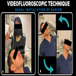
- A 5 -10ml syringe is filled with thick consistency barium and connected to a pediatric feeding tube, inserted about 6- 8 cm beyond external nares into nasal cavity, 3-5 cc of the material is introduced each nostril with the head of the patient hyperextended. (We recommend the patient blows both sides of the nose to expel retained mucus). (See Figure 7).
- The patient must be encouraged to sniff in through his nose to precipitate the posterior flow of the barium. Tilting of the head is recommended for 1 min to let the barium to impregnate the velum and nasopharyngeal walls.
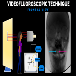
- Barium coating tends to last just a few minutes in the lateral pharyngeal walls. Starting with frontal view during speech is recommended. (See Figures 8 and 9).
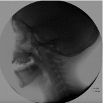
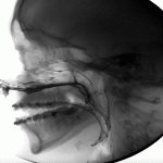
- Lateral views during speech must be taken. It´s important that the head is not tilted o rotated. Oblique views can be used in asymmetric velopharyngeal movements. (See. Figures 10 and 11).
- Once the barium is instilled nasally, the examination must be performed rapidly. Recommended examination time is 40 seconds.
- Examination is finished.
3.- RADIOLOGICAL REPORT.
-Frontal View.
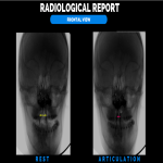
- Distance between lateral pharyngeal walls at rest.
- Distance between lateral pharyngeal walls during articulation. See (Figure 12)
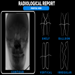
- Contour: Refers to the vertical movement of each lateral pharyngeal wall, these movements can be referred as, shelf, balloon, vertical and irregular. See (Figure 13).
- Direction of the vector of movement can be medial, superomedial, and outward.
- Symmetry or asymmetry in the lateral pharyngeal wall closure during phonation, contour, and direction of the vector of movement should be described.
-Lateral View.
- Distance between the tip of the uvula and the hard palate at rest.
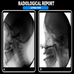
- Percentage of velopharyngeal closure gap (or size of the gap). See (figure 14).
- Direction of velar movement: In the plane of the hard palate and can be superior, posterior, posterosuperior, and tip-hinge (the velar tip elevates to touch the pharyngeal posterior wall
- Presence or absence of Passavant’s ridge See (figure 15).
- Direction of Passavant’s ridge if present: anterior or anterosuperior.
- Presence or absence of Lymphoid tissue.


