Learning objectives
- To review spinal cord anatomy as well as its vascularization.
- To present the typical clinical and radiological patterns of spinal cord infarctions.
- To learn how to differentiate it from other potentially curable pathologies with similar clinical presentation.
Background
Although spinal infarctions represent less than 1% of the vascular pathologies of the central nervous system, they can leave serious sequelae that significantly decrease the patient's quality of life.
MRI examination that include diffusion-weighted sequences is the “gold standard” test in patients with clinical suspicion of spinal cord ischaemia; that also allow to make adequate differential diagnosis.
Findings and procedure details
Anatomy:
The spinal cord is part of the central nervous system that extends from medulla oblongata to lumbar region (level of vertebral body L1-L2).
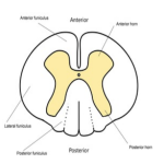
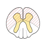
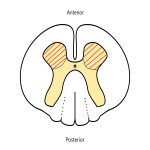
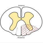
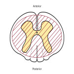
It is made by H-shaped grey matter that forms the most inner part of the spinal cord and white matter around it. Grey matter forms anterior and posterior horns and split white matter into funiculus (anterior, medial and posterior) (Fig. 1) [Fig 1].
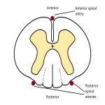
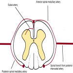
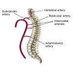
The spinal cord is irrigated by three arteries: two posterior spinal arteries (PSAs) and one anterior spinal artery...
Conclusion
The radiological assessment of the infarct extension and the affected territory can predict the severity of the sequelae and their future management.
It is of great importance to make adequate differential diagnosis with other potentially curable pathologies that present a similar clinical picture.
Personal information and conflict of interest
M. HERNANI ÁLVAREZ:
Nothing to disclose
F. J. Gwiazdowski:
Nothing to disclose
A. Mora Jurado:
Nothing to disclose
References
Masson C, Pruvo J, Meder J et al. Spinal Cord Infarction: Clinical and Magnetic Resonance Imaging Findings and Short Term Outcome. J Neurol Neurosurg Psychiatry. 2004;75(10):1431-5.
Thurnher M & Bammer R. Diffusion-Weighted MR Imaging (DWI) in Spinal Cord Ischemia. Neuroradiology. 2006;48(11):795-801.
Vargas M, Gariani J, Sztajzel R et al. Spinal Cord Ischemia: Practical Imaging Tips, Pearls, and Pitfalls. AJNR Am J Neuroradiol. 2015;36(5):825-30.
Novy J, Carruzzo A, Maeder P, Bogousslavsky J. Spinal cord ischemia: clinical and imaging patterns, pathogenesis, and outcomes in 27 patients. Arch...


