Keywords:
Cardiac, MR, Technical aspects, Blood
Authors:
R. G. Chelu1, A. Hsiao2, A. Coenen1, M. L. Dijkshoorn1, J. Roos-Hesselink1, G. P. Krestin1, K. Nieman1; 1Rotterdam/NL, 2San Diego, CA/US
Methods and Materials
Methods
Between November 2015 and February 2016,
we prospectively included 10 consecutive adult patients (4 females,
mean age 35 yo) known with bicuspid aortic valve.
The MR and CT scan were performed in the same day.
The 4D flow raw data sets were uploaded to a dedicated web-based software application (Arterys Inc.,
San Francisco,
CA,
USA).
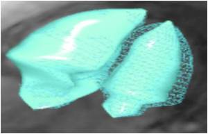
Images were reconstructed in 20 cardiac temporal phases separately with a compressed sensing algorithm.
The end-diastolic,
end-systolic and stroke volumes and ejection fractions were measured by CMR 4D flow.

Fig. 1
Functional CT data acquisition was performed on a 3rd generation dual-source CT (Definition,
Siemens Medical Solutions,
Germany).
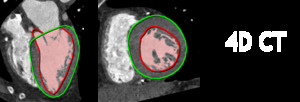
Imeges were also reconstructed during 20 cardiac phases and measurements were performed in end-diastolic and end-systolic phases,
using for post-processing SyngoVia (Siemens Medical Solutions,
Germany).

Fig. 2
.
In both modalities papillary muscles were included in the left ventricle cavity.


