ESSR 2017 / P-0209
Adult Hip Radiography: Lines and angles
Congress:
ESSR 2017
Poster Number:
P-0209
Type:
Educational Poster
Keywords:
Education and training, Education, Conventional radiography, Musculoskeletal bone, Bones
Authors:
A. L. Proenca, A. P. Caetano, L. Bogalho; Lisbon/PT
DOI:
10.1594/essr2017/P-0209

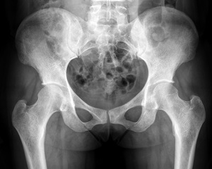
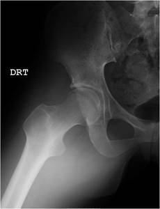
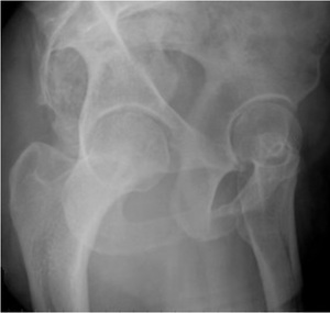
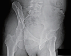
Fig. 1:
Anteroposterior view of the pelvis
")
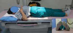
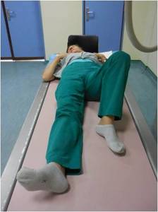
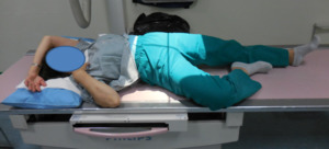
Fig. 2:
AP Positioning
- Patient supine on the x-ray table.
- Both lower...

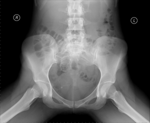
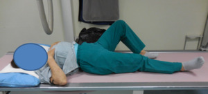
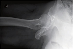
Fig. 3:
Frog-leg lateral view
, affected knee flexed (30-40º), thighs abducted (45º) and externally rotated.")
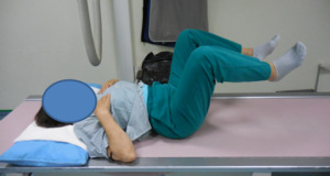
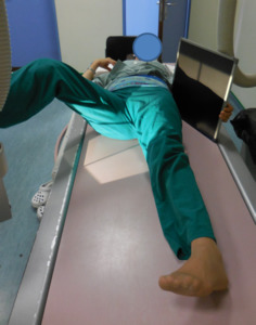
Fig. 4:
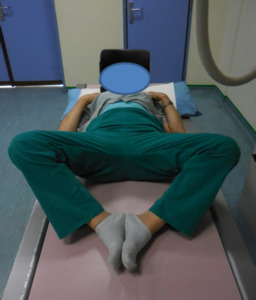
Frog-leg lateral view Positioning
- Patient supine on the x-ray table.
-...

Fig. 5:
Dunn view at 45º

Fig. 6:
Dunn view at 45º Positioning:
- Patient supine on the x-ray table.
-...

Fig. 7:
Dunn view at 45º

Fig. 8:
Dunn view at 90º Positioning
- Patient supine on the x-ray table.
- Pelvis...

Fig. 9:
Cross-table lateral view

Fig. 10:
Cross-table lateral view Positioning
- Patient supine on the x-ray table.
-...

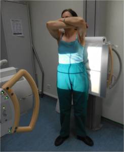
Fig. 11:
False-Profile view

Fig. 12:
False-Profile view Positioning
- Patient in orthostatic position.
-...
 References: 2015 by Korean Hip Society")
Fig. 13:
Judet view. Right posterior oblique (downside)
.")
Fig. 14:
Image 13: Judet view Positioning
- Patient in a 45° oblique position.
-...