ESSR 2017 / P-0219
Normal radiologic findings and detection of complications in Total Hip Arthroplasty
Congress:
ESSR 2017
Poster Number:
P-0219
Type:
Educational Poster
Keywords:
Prostheses, Surgery, Conventional radiography, Musculoskeletal joint, Musculoskeletal bone
Authors:
A. L. Proenca, A. P. Caetano, L. Bogalho; Lisbon/PT
DOI:
10.1594/essr2017/P-0219

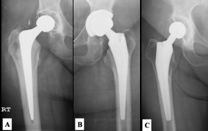
Fig. 1
, non-cemented(B) and hybrid (C - cemented acetabular component and non-cemented femoral component) THA.")
Fig. 2:
Cemented (A), non-cemented(B) and hybrid (C - cemented acetabular component and...

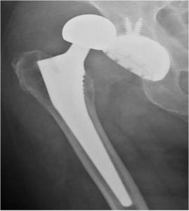
Fig. 3:
Visible acetabular and femoral components in a femoral head dislocation.