Keywords:
Inflammation, Infection, Genetic defects, Education, Complications, Plain radiographic studies, CT, Conventional radiography, Thorax, Respiratory system
Authors:
P. Patel, I. Karafotias, S. D. Tran, N. Mulholland, G. Yusuf, K. Stefanidis; London/UK
DOI:
10.26044/esti2019/P-0024
Background
Primary humoral immunodeficiency constitutes the vast majority of all immunodeficiencies,
characterised by insufficient or impaired antibody production which,
can lead to increased susceptibility to infections.
The clinical presentation,
complications and the radiological appearances depend on the type of defects with a great variability between genotypes and phenotypes (Table 1 and 2).
Common variable immunodeficiency (CVID) and X-linked agammaglobulinaemia (XLA) are the commonest types of humoral immunodeficiency presenting with respiratory symptoms and a variety of pulmonary imaging findings.
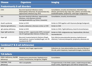
Table 1: Correlation of the subtypes of the various primary immunodeficiencies with the most common pathogens and description of the possible radiology manifestations.
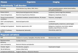
Table 2: Correlation of the subtypes of the various primary immunodeficiencies with the most common pathogens and description of the possible radiology manifestations.
