We did a retrospective study over a period of 11 months in children presenting to the emergency department of our hospital with history of blunt trauma to abdomen. Patients between age of 2 to 18 years who were referred to our department for CECT examination were included in our study.
Initial examination was Focused Assessment with Sonography in Trauma (FAST) followed by biphasic/triphasic contrast enhanced Computed Tomography. All scans were performed with Philips 64 slice scanner.
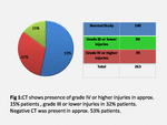
Severity of injury in CT was assessed on American Association for the Surgery of Trauma (AAST) grading for solid organ injury and were categorized as(Fig. 1):
1. Grade IV or higher injury in one or more solid organs(s)
2. Grade III or lower injury in one or more solid organ(s)
3. Normal/Negative CT study
Correlation of the following five variables was done with the CT findings:
1.Age of the child
2.Focused abdominal sonography in trauma (FAST) status.
3.Mechanism of injury
4.Presenting complaints and clinical features (hypotension, tachycardia etc).
5.Other radiological studies (Chest, abdominal or extremity x rays).
Age: We divided the patients into two groups, i.e., less than 10 years of age and more than 10 years of age, then looked at the prevalence of category of injury in each of them.
(Fig. 2)
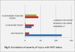
Focused abdominal sonography in trauma (FAST) status: We tried to quantify the FAST status as: (Fig. 3)
1. Negative FAST examination
2. Fluid in one pocket (e.g. pelvis only)
3. Fluid in more than one pocket.
Pockets refer to hepato-renal, spleno-renal, pelvic, pericardial and pleural cavities.
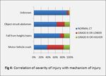
Mechanism of injury: Most commonly encountered mechanisms were motor vehicle crash, fall from height/stairs, object striking abdomen (e.g. bicycle handle) and unknown. (Fig. 4)
Presenting complaints and clinical features: As it forms a part of assessment in Royal College of Radiologist paediatric trauma protocol, [9] presenting features like lap or seat belt injury, abdominal wall ecchymosis, abdominal distention and nasogastric or per-rectal bleed were included. Clinical features like clinical evidence of hypotension and or unexplained tachycardia were considered.
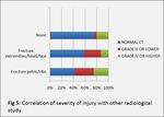
Other radiological studies: Most of the patients underwent conventional radiological studies like X-ray chest, abdomen or extremities depending on presenting features. (Fig. 5)
All five variables were statically analysed and p-values ( Fig. 6 ) were derived for age, mechanism of injury, presenting complains, clinical features and other radiological investigations, while parameters like sensitivity and specificity were determined for FAST status.
Except for presenting features, all other variables well correlated with severity of injury with p-values <0.05, while for FAST status, sensitivity was found to be approx. 89.4%, specificity approx. 85% and negative predictive value approx. 90%. ( Fig. 7 )


