Congress:
EuroSafe Imaging 2020
Keywords:
Performed at one institution, Not applicable, Cancer, Radiation effects, MR, CT, Radioprotection / Radiation dose, Head and neck, Head and Neck
Authors:
R. T. Le, F. Behzadi, P. Fiester, J. Patel, R. Dagan, D. Rao
DOI:
10.26044/esi2020/ESI-08297
Description of activity and work performed
Both the World Health Organization (WHO) and Radiation Therapy Oncology Group (RTOG) have classified RIOM into a set of four major clinical stages [Table 1] [3,4,5]. The RTOG grading symptom is utilized more in the clinical setting [4].
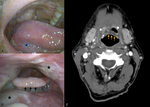
Macroscopic signs of RIOM [Fig. 2] appear on non-keratinized surfaces (e.g. tongue, buccal mucosa, soft palate), initially as erythema, and can progress to desquamation approximately one month after the beginning of radiotherapy. Corresponding radiographic findings would show mucosal edema and swelling of the epiglottis. Patients may report dysphagia, oral pain, and difficulty swallowing, which may correlate with more severe mucosal swelling and a more severe mucositis.
Repeated radiotherapy exposure can increase the severity of mucositis, leading to more organized RIOM (Grade III) or oro-pharyngeal necrosis (Grade IV). If a patient develops Grade III mucositis during radiotherapy, it is recommended to cease radiotherapy to minimize further complications.
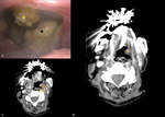
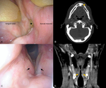
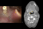
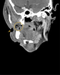
In advanced stages (Grade IV and V), a patient will have more extensive erythema or ulceration and may be unable to tolerate oral intake. Examples of wound breakdown include mucosal wall necrosis [ Fig. 3], osteonecrosis [Fig. 4, Fig. 5], and/or fistula formation [Fig. 6] requiring a combination of antibiotic treatment, hyperbaric oxygen, and/or surgical intervention.


