Objectives
1. What type of imaging is used to investigate trauma patients?
2. Are the scans indicated based on the presentation and clinical examination?
3. Are CTs being performed and reported within the recommended times frames?
4. How many of these patients have significant findings?
Method
Trauma patients who attended the emergency department between 20/07/2019 to 20/08/2019 were identified using CDN Radiology Information System (CRIS). Time of arrival, clinical information, scan type and time, time report verified and significant findings were recorded.
Method
Trauma patients who attended the emergency department between 20/07/2019 to 20/08/2019 were identified using CDN Radiology Information System (CRIS). Time of arrival, clinical information, scan type and time, time report verified and significant findings were recorded.
Results
1. CT head was the most common imaging modality, representing 82% of total CT scans performed. The majority of CT Heads were indicated (83%). Out of 361 CT head scans, 4 showed an acute fracture, 16 showed intracranial haemorrhage, 1 showed a scalp haematoma and 4 showed incidental tumours. 53% of scans indicated within an hour of presentation were performed within the target time. Of those that did not meet the 1 hour target, 84% were performed within the second hour. 98% of CT heads indicated within an 8 hour window were performed within this target window. Reports were issued within 1 hour in 81% of cases. There was little difference in reporting times between in-house radiologists and scans which were out sourced externally for reporting.
2. 87% of c-spine CTs were indicated. Out of 47 CT c-spines 2 had positive findings of undisplaced fractures.
3. 66% of CT thorax/abdomen/pelvis scans did not appear to be indicated according to the request.
4. Only 5% of patients had an x-ray prior to a CT of the c-spine, thorax, abdomen or pelvis scan. 1 of these x-rays showed an abnormal finding of a haemothorax. Out of the 80 scans performed 12.5% had positive findings (7 rib/vertebral fractures, 1 pleural effusion, 1 haemothorax and 1 splenic rupture).
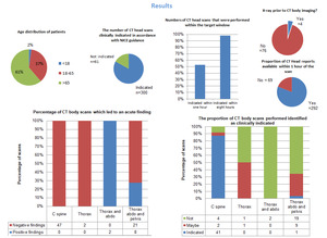
Fig. 1
