Learning objectives
This paper presents a six-year experience in an Australian and New Zealand setting of applying computer algorithms to automate the analysis of MRI images of the liver for the quantification of iron and fibrosis,
and the insights and learnings achieved over this period to transform these algorithms into a regulated medical device.
Background
The following terms are used either throughout the paper or are essential background knowledge to an understanding of the methodologies relating to image processing and computer based artificial intelligence:
Machine Learning is the use of algorithms to allow computers to learn models from data without being explicitly programmed.
Support Vector Machines – use supervised learning methods (see below) for classification.
The SVM is presented with examples of images that have been classified into one of two categories.
The SVM builds a model that predicts whether...
Imaging findings OR Procedure details
Part One: Support Vector Machine Learning - Liver Fibrosis
In 2012,
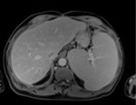
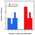
we applied support vector machine learning (SVM) to a cohort of 28 patients with hepatitis C to assess textural differences in MRI images [2] to build a predictive model for categorising the patients’ Ishak fibrosis scores [3].
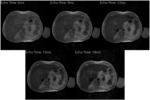
Figure 1 shows an example liver image used in the study.
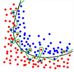
The assumptions and limitations of the modelling of small patient numbers,
that include cross-validation and overfitting,
are presented.
The small subject population was divided into both...
Conclusion
Other things to consider when building a neural network:
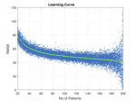
This paper reports a six-year experience applying machine learning models to small and large image datasets.
The key elements that have resulted in an acceptable model are the growth in computer processing power,
access to advanced machine learning algorithm libraries,
and access to large volumes of consistently acquired and well labelled data.
Although the majority of the analysis in this paper has focussed on the application of computer algorithms to automate the analysis of MR images...
Personal information
Martin Blake: MBBS,
FRANZCR,
FAANMS,
MBA,
FAICD
Martin is a Radiologist and Nuclear Medicine Specialist,
with 30 years medical imaging experience and has been a partner of Perth Radiological Clinic since 1997 and Chairman since 2009.
He has also been a non-executive director of Resonance Health (ASX:RHT) since 2007 and Chairman since 2012.
Tony Cooper: BSc (Hons),
MS (Statistics)
Tony is a Data Scientist and is a founder of Precision AI which provides artificial intelligence solutions to European and Australasian industry.
He has a distinguished...
References
Brownlee J.
Supervised and Unsupervised Machine Learning Algorithms,https://machinelearningmastery.com/supervised-and-unsupervised-machine-learning-algorithms/
House M.J.,
Bangma S.J.,
Thomas M.,
Gan E.K.,
Ayonrinde O.T.,
Adams L.A.,
Olynyk J.K.,
St.
Pierre T.G.
(2015) Texture-based classification of liver fibrosis using MRI.
Journal of Magnetic Resonance Imaging 41: 322-328.
St Pierre T.G.,
House M.J.,
Mian A.,
Bangma S.,
Burgess G.,
Standish R.A.,
Casey S.,
Hornsey E.,
Angus P.W.
(2015) Machine-learned Image Analysis Models for Classifying Liver Fibrosis Stage from Magnetic Resonance Images.
Hepatology 62: 607A.
Pan S.J.
and Yanq Q.,
"A Survey on Transfer...


