
ECR 2014 / C-1088
Intracranial haemorrhage on phase images of SWI (susceptibility weighted image)
This poster is published under an open license. Please read the disclaimer for further details.
Congress:
ECR 2014
Poster Number:
C-1088
Type:
Scientific Exhibit
Keywords:
Neuroradiology brain, MR, CT, Outcomes analysis, Haemorrhage
Authors:
Y. J. LEE, H. S. Choi, B.-Y. Kim, J. Jang, S. L. Jung, K. J. Ahn, B. S. Kim; Seoul/KR
DOI:
10.1594/ecr2014/C-1088

Fig. 1:
Pattern of hemorrhage in phase image.
 on CT and MRI with different values on phase image on 3 different stages.
A, CT and MRI show acute SDH along the right cerebral convexity. Phase image shows heterogeneous mixed bright and dark value(arrows). B, CT and MRI show subacute SDH along left cerebral convexity. Phase image shows heterogeneous mixed bright and dark value(arrow).
C, CT and MRI show late subacute to chronic stage of SDH along the both cerebral convexity(arrows). SWI shows iso signal(arrows). Phase image shows homogeneous gray value(arrows).")
Fig. 2:
Subdural hemorrhage(SDH) on CT and MRI with different values on phase image on...
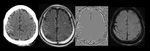
Fig. 3:
Sixty-one year old male patient with subarachnoid hemorrhage(SAH).
CT images...

Fig. 4:
All parenchymal hemorrhages more than 5-mm showed aliasing pattern on phase...