Purpose
Arrhythmogenic right ventricular cardiomyopathy (ARVC) is a major cause of sudden cardiac death and heart failure (1).
It ischaracterized by fibro-fatty replacement of normal myocardial tissue,
predominantly on the sub-epicardial layers of the right ventricule (RV) (2).
The presence of myocardial fat can be assessed onMDCT orMRI but this imaging feature is not part of recommendations to diagnose ARVC(3)because it lacks specificity and reproducibility (4).
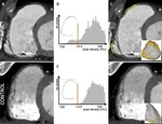
The aim of this study was to introduce an automatic method for the quantification of myocardial fat in the RV...
Methods and Materials
Population
36 ARVC patients were compared to 36 age and sex-matched subjects with no structural heart disease (control group),
as well as to 36 patients with ischemic cardiomyopathy (ischemic group).
MDCT acquisition and segmentationof RV fat
Patients underwent contrast-enhanced ECG-gated cardiac MDCT with a biphasic injection protocol to optimize RV enhancement.
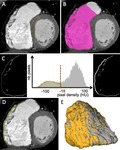
Image segmentation and fat quantification were performed using an automatic method as shown in Figure 1.The extent of fat was expressed in % of RV fre wall.
Registration of fat distribution on RV template...
Results
Patients characteristics
Characteristics of ARVC patients (46±15yrs,
7 women) are shown in Table 1.
All had definite ARVC diagnosis according to Task Force criteria.
In the control group(47±15yrs,
7 women),
all patients were referred at MDCT for the assessment of ascending aorta,
and had no history of palpitaion,
syncope,
or structural heart disease.
In the ischemic group(62±12yrs,
4 women),
all patients had history of myocardial infarction.
Reproducibility of MDCT measurements
The acquisition and processing of MDCT images were feasible in all patients.
Intra and inter-observer...
Conclusion
Automated quantification of fat in RV free wall from MDCT images is feasible and reproducible.
The diagnostic performance of the method in ARVC is superior to that of RV volume,
yielding similar specificity and markedly improved sensitivity.
Besides global quantification,
the method also allows for three-dimensional mapping of fat over the RV free wall,
demonstrating that basal lateral fat is the most specific of ARVC.
References
Sen-Chowdhry S,
Syrris P,
Ward D,
Asimaki A,
Sevdalis E,
McKenna WJ.
Clinical and genetic characterization of families with arrhythmogenic right ventricular dysplasia/cardiomyopathy provides novel insights into patterns of disease expression.
Circulation 2007;115:1710 –1720
Marcus FI,
Fontaine GH,
Guiraudon G,
et al.
Right ventricular dysplasia: a report of 24 adult cases.
Circulation 1982;65:384-98.
Marcus FI,
McKenna WJ,
Sherrill D,
et al.
Diagnosis of arrhythmogenic right ventricular cardiomyopathy/dysplasia: proposed modification of the task force criteria.
Circulation 2010;121:1533-41.
Bomma C,
Rutberg J,
Tandri H,
et al.
Misdiagnosis...


