Introduction
Congenital cardiovascular anomalies (CCVAs) remain considerable health problem in pediatric population.
The reported incidence is approximately 6-8 per 1000 at birth,1 but after popularization of colour Doppler echocardiography that makes it possible to detect and characterize asymptomatic lesions,
most investigators report higher number of its incidence; 2 as in 2007 Andrew Crean reported that children with congenital heart disease account one in every 145 live births in the UK.3
CCVAs may present as a sole abnormality of stenosis,
obstruction,
defect or connection problems or as a complex combination of these features.4
Radiology plays an important role in initial detection of congenital cardiovascular abnormalities as well as in evaluation of post treatment conditions.
Imaging can give structural and functional information about cardiovascular system,
help to plan management,
evaluate the results of intervention and predict longtime outcomes.1
The improvement in early detection and management of CCVAs have resulted in increased survival in pediatric population.5
Today various imaging modalities are utilized in detection and follow up of congenital cardiovascular anomalies and the trend in choosing the appropriate imaging modality is towards obtaining: more accurate morphological and/or functional information with non/less invasive techniques and reduced/no radiation exposure.
Radiological approach of children with CCVAs
Some CCVAs can be easily diagnosed by one modality but,
sometimes patients may have complex anomalies of the thoracic vasculature as none of the available imaging modalities can answer all the questions by alone,
so for precise and detailed depiction of the cardiovascular morphology and function,
a multi-modality approach is required in a complementary fashion simultaneous to the clinical context and primary findings to reach the accurate diagnosis and plan treatment.1
The important and frequently performed radiological examination is still chest radiography.2 However of low sensitivity in precise detection of CCVAs,
this easily available technique can give clues of suspected CCVAs and offer valuable information in order to narrow the differential diagnosis to few likely conditions.6
Echocardiography is the first-line imaging technique for establishing the diagnosis and follow up in most patients with congenital cardiovascular anomalies.
7 If echocardiography could not answer all the raisin questions,
then cardiac magnetic resonance imaging (CMRI) and multi detector computed tomography (MDCT) angiography can be the next imaging modalities of choice before proceeding to more invasive technique: angiocardiography.
Invasive studies can still be nessecery in more complex conditions.2 Nuclear imaging is used in selected circumstances.8
Ashwin Prakash presented a general diagnostic algorithm for choosing the appropriate imaging technique according to the patient’s clinical context,
primary imaging findings and subsequent specific diagnostic questions.8 This algorithm is copied in (Figure 1).
(However this algorithm is not specifically made for pediatric patients)
1- Chest radiograph
Chest radiography is still the most important and most frequently performed radiological examination.2 With development of modern imaging techniques,
the role of plain chest radiography in evaluation of congenital cardiovascular anomalies has largely been relegated to only of historical and academic interest,
however it can give valuable information and narrow the differential diagnosis to few likely conditions.6 A number of plain chest radiography signs of congenital cardiovascular abnormalities are well described in literature that are of significant clinical importance.9 (Figures 2 and 3)
2- Echocardiography
Echocardiography is the first-line imaging technique for establishing the diagnosis and follow up in most patients with congenital cardiovascular anomalies7 and should always be performed before proceeding to other modalities.8 (Figure 4)
This non-invasive,
portable,
radiation free,
widely availability and unexpansive modality gives high resolution anatomical and functional information about cardiovascular system and has a high accuracy in detection of intra cardiac lesions.1,7
Usually there is no need for sedation or anesthesia in children undergoing echocardiography.
For co-operative patients with appropriate acoustic windows,
echocardiography by alone can define diagnosis,
guide management and evaluate prognosis,1 but if the acoustic windows are poor,
particularly for extra-cardiac vascular structures it will fail to give all the needed information.
In a study in 2013 Al-Azzazy Mohamad Zakaryia has concluded that the overall sensitivity of Doppler echocardiography in detection of intra cardiac defects is 100%,
while for extra-cardiac aortic anomalies its sensitivity is 92%.10
The optimal echocardiographic examination should fulfill the following three criteria:
1. High enough quality exam to ensure that the clinical condition being managed is well understood.
2. Free from ambiguities about the severity of the condition.
3. Complete,
to ensure that there are no clinically relevant unappreciated cardiovascular conditions.
If any of these criteria are not confidently fulfilled,
then echocardiography cannot be the only source of imaging information on which management should be based and there is need to proceed to further imaging techniques.11
The current literature demonstrates the diagnostic accuracy of echocardiography in evaluation of congenital heart disease around 80% and in the rest of 20%,
the defect is hiding in the dead area of echocardiography.12
The major limitations of echocardiography are: operator dependence,
limited acoustic window,
poorer spatial resolution and inappropriate access for evaluation of extra-cardiac lesions.7 Assessment of an entire congenital repair is often suboptimal and neither the tracheobronchial structures nor the lung parenchyma can be evaluated by this technique.
Furthermore echocardiography is not great in evaluation of coronary arteries,
quantitative assessments of valvular dysfunction,
ventricular volumetrics,
and myocardial masses.11
3- Cardiac Computed Tomography Angiography (cCTA)
Cardiac CT Angiography is an important non-invasive and rapid imaging technique providing excellent anatomic information with high spatial resolution and powerful three dimensional post-processing image reconstruction about cardiovascular system.7 (Figure 5)
It can accurately depict complex cardiovascular anatomic features both before and after surgery as well as a variety of post treatment complications.13
As previously mentioned that echocardiography is excellent in delineation of intra cardiac lesions,
non-ECG gated acquisition cCTA is usually used for evaluation of extra cardiac vascular structures while the ECG-gated acquisition cCTA is used for evaluation of coronary artery.7
Al-Azzazy Mohamad Zakaryia reported the overall sensitivity of the MDCT angiography in detection of extra-cardiac aortic anomalies as 100% compared to 92% of Doppler echocardiography while for intra cardiac defects sensitivity of MDCT angiography was 85% compared to 100% sensitivity of Doppler echocardiography.10
Cardiac CTA is appropriate for evaluation of children with pacemakers,
internal cardiac defibrillators or aneurismal clips in whom MRI is contraindicated.7
Due to its short examination time,
CT is also applicable in emergency settings for critically ill child who cannot tolerate MR imaging.14 Usually there is no need for general anesthesia.
Despite above mentioned advantages,
cCTA is not the modality of choice for evaluation of ventricular and valvular function,
blood flow and shunt quantification as well as myocardial viability.7
The most important issue regarding CCTA is the radiation exposure and the trend is how to keep the radiation dose as low as reasonably practicable (ALARP) with the balance of adequately interpretable image quality.
The newer generation (256 to 320/640-slice) scanners reduce the scan time to less than a second and consequently the radiation exposure to a minimal dose.7
4- Cardiovascular Magnetic Resonance Imaging (cMRI)
Cardiac MR and CT have revolutionized the management CCVAs.
The ability of cMRI in providing high resolution,
three dimensional cardiac imaging with accessibility to hemodynamic evaluation made it an appropriate,
noninvasive,
radiation free alternative for both echocardiography and cardiac catheterization.1 Also cCTA is not the modality of choice for evaluation of ventricular and Valvular function,
blood flow and shunt quantification as well as myocardial viability,
so these can be imaged by radiation-free cMRI.
Cardiac MRI is increasingly used in neonates and infants for the initial investigation of CCVAs or as follow-up after surgery or catheter-guided intervention.10 Christian J.
Kellenberger has listed the common clinical indications for cardiovascular MR imaging in neonates and infants with congenital heart disease as: investigation of thoracic vasculature in complex CCVAs,
quantification of ventricular volumes,
Follow-up after surgery or intervention and evaluation of primary cardiac tumors.15
Also Cardiac MRI is ideally suited to post-surgical follow up examinations in this relatively radiosensitive population as repeated scans may be needed. CT with is best reserved for those in whom CMR is not possible.3
The need for deeper sedation or general anaesthesia,
less availability,
higher cost,
longer examination time,
related lower spatial resolution are the disadvantages of cMRI compared to ECG gated cCTA.
Suvipaporn Siripornpitak made a comparison between cCTA and cMRI in evaluation of congenital heart disease which is copied in (table 1)
5- Cardiac catheterization:
Cardiac catheterization had been the reference standard for the investigation of CCVAs many years,3 but due to its invasive nature (may cause death in up to 1% of neonates with complex CHD16) and development in noninvasive imaging modalities,
it is currently largely replaced by advanced non-invasive imaging techniques and is now uncommon for newborns to undergo catheterization for purely diagnostic purposes.7
In addition to owing invasive technique,
relatively high radiation dose and need for large amount of iodinated contrast injection,
catheter cardio angiography is limited by the overlapping of adjacent vascular structures and resultant problem in demonstrating systemic and pulmonary vascular systems simultaneously.4
Echocardiography is now usually preferred as the primary diagnostic modality whereas cardiac MRI and CT mostly perform the advanced functions needed.
Cardiac catheterization is currently reserved for hemodynamic assessment,
in cases with pulmonary hypertension and complex Congenital cardiovascular diseases in whom the data regarding pulmonary vascular resistance,
oxygen saturation and chamber pressure are essential for surgical planning,
and in whom interventional treatment is necessary.17 (Figure 7)
6- Nuclear Scintigraphy
Nuclear imaging is used in selected circumstances.
Positron emission tomography (PET) can be used to evaluate myocardial viability and metabolism.
Lung perfusion with 99mTclabeled macroaggregated albumin can be used to determine differential flow to each lung,
in determining the physiological significance of pulmonary artery or vein stenosis.
However due to its relatively low spatial resolution and exposure to ionizing radiation,
nuclear scintigraphy plays a limited role in the pediatric patients.8

: 112-125.")
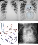
 Chest radiograph obtained in an infant with a right-sided aortic arch (b the same as a with a superimposed drawing) shows the characteristic boot-shaped sign produced by upturning of the cardiac apex because of right ventricular hypertrophy and by the concavity of the main pulmonary artery. References: Ferguson, Emma C., et al. "Classic imaging signs of congenital cardiovascular abnormalities." Radiographics 27.5 (2007): 1323-1334.")
 and overriding aorta in tetralogy of Fallot
(RV, right ventricle; LV, left ventricle; LA, left atrium; AO, aorta). References: Plappert, Ted, and Martin G. St John Sutton. The Echocardiographers' Guide. CRC Press, 2006.")
: VRT and MPR reconstructed coronal CT images showing Left sided vertical vein draining to left brachiocephalic vein and subsequently joins right brachiocephalic vein to drain into the superior vena cava.
C: Axial MPR CT image at atrial level demonstrating an arterial septal defect between dilataed right and hypoplastic left atrium.
D: coronal MPR reconstructed CT image showing Prominent right and left pulmonary veins draining to the vertical vein posterior to the heart instead of draining to the left atrium.")
. References: Prakash, Ashwin, Andrew J. Powell, and Tal Geva. "Multimodality noninvasive imaging for assessment of congenital heart disease." Circulation: Cardiovascular Imaging 3.1 (2010): 112-125.")
: Left anterior oblique VR (a) and subvolume MIP (b) MR angiographic images reveal restenosis of the aortic arch, including the origin of the left subclavian artery (arrow).
On the basis of the MR angiographic findings, it was decided to perform
catheter-guided dilation of the aorta and subclavian artery.
(c, d) Left anterior oblique angiograms help confirm the degree of stenosis seen at MR angiography and show a good result after dilation. References: Kellenberger, Christian J., Shi-Joon Yoo, and Emanuela R. Valsangiacomo Büchel. "Cardiovascular MR Imaging in Neonates and Infants with Congenital Heart Disease 1." Radiographics 27.1 (2007): 5-18.")
