ECR 2008 / C-176
Myocardial bridging and related coronary atherosclerotic burden by 64-slice CT coronary angiography
Congress:
ECR 2008
Poster Number:
C-176
Type:
Scientific Exhibit
Keywords:
Authors:
L. La Grutta1, G. Runza1, V. Alaimo1, L. Damiani1, F. Alberghina1, G. Lo Re1, F. Cademartiri2, M. Midiri1, R. Lagalla1; 1Palermo/IT, 2Parma/IT
DOI:
10.1594/ecr2008/C-176
DOI-Link:

Fig. 1:
Myocardial bridge

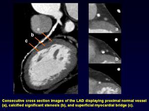
Fig. 2:
Superficial myocardial bridge with associated proximal significant stenosis

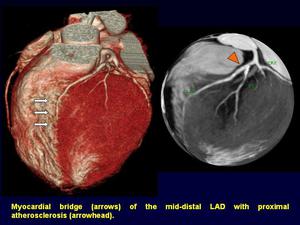
Fig. 3:
Superficial myocardial bridge of the distal LAD and proximal associated mixed...