Keywords:
Musculoskeletal spine, Musculoskeletal system
Authors:
W. G. Bugg, M. Lewis, A. Juette, J. Cahir, A. P. Toms; Norwich/UK
DOI:
10.1594/ecr2010/C-2414
Methods and Materials
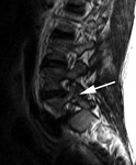
This is a retrospective case control study. Patients with pars interarticularis fractures demonstrated on MRI (Figure 1) were identified from Radiologists’ databases and from free text searches in RIS. Data was collected from our PACS archive for the years 2002 to 2009. Inclusion criteria included all cases with bilateral L5 pars interarticularis fractures with an accompanying standing lateral radiograph of the lumbar spine. All examinations were reviewed independently by two musculoskeletal consultant radiologists. MR examinations were excluded if there were any abnormalities, other than the pars fracture, including segmentation anomalies, degenerative disc disease, facet joint osteoarthrosis and spondylolisthesis greater than grade 1. Any cases with signal loss in the nucleus pulposus on T2W sagittal images or any vertebral translation anteroposteriorly were excluded.
Age and sex matched controls were found from PACS for each case. The control cases were assessed independently by the same two musculoskeletal consultant radiologists. Cases were again excluded if evidence of segmentation anomalies, or other MRI changes associated with back pain were identified which could affect the angle of lordosis. If a concern was raised over the suitability of a case of spondylolysis or control case by either observer it was excluded from the study.
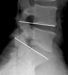
Standing lateral lumbar spine radiographs for the both the spondylolysis and control cases were assessed for the angle of lumbar lordosis. The lumbar spine radiograph most recent to the lumbar MRI was selected for inclusion. A constrained Cobb angle measurement [11] was taken between the inferior endplate of the L4 vertebra and the superior endplate of the S1 vertebra using the 2-line technique [12] on a high resolution 2K PACS workstation (Barco, x,Belgium) (Figure 2). Line placement was performed manually by two independent observers.
Descriptive statistics of the angles of lordosis and a Student-T test for the difference in the means between the two groups were calculated. Inter-rater reliability was measured using interclass correlation coefficients (MedCalc v2.0).


