The Tumor-Node-Metastasis (TNM) staging system for non-small cell lung cancer (NSCLC,
accounting for the 87% of all lung cancers) is an internationally accepted system used to determine the disease stage.
This disease stage is a measure of the extent of disease,
which is used to guide the clinical-diagnostic stage,
surgical-pathologic stage,
retreatment stage,
autopsy stage and prognosis.
The TNM staging system for NSCLC categorizes tumors on the basis of primary tumor characteristics (T),
the presence or absence of regional lymph node involvement (N),
and the presence or absence of distant metastases (M).
The overall stage of the tumor (stage I through IV) is determined by the combination of T,
N,
and M grades.
The clinical-diagnostic stage is based upon medical history,
physical examination,
laboratory testing,
radiologic testing,
tissue sampling,
and any other investigation undertaken prior to primary therapy.
It is assigned the prefix c (eg,
cT3N2M0).
A limitation of clinical-diagnostic staging is that the stage is sometimes related to the intensity of the evaluation.
The surgical-pathologic stage is based on the clinical-diagnostic stage plus histopathologic data from the resected tumor.
It provides confirmation of the T descriptor,
N descriptor,
and histologic type.
In addition,
it takes into account the histologic grade,
resection margins,
and presence or absence of lymphovascular invasion.
The surgical-pathologic stage is assigned the prefix p (eg,
pT3N2M0).
A retreatment stage is assigned if there is recurrence of disease and a new treatment program is planned.
An autopsy stage is recorded when a patient dies and has a postmortem examination performed.
The suffix "X" is attached (eg,
TX,
NX,
or MX) if the extent of disease cannot be assessed for any of these features.
This staging system is based upon a retrospective analysis of survival in diverse samples of patients representing all stages of disease.
It reflects the clinical evaluation methods and treatments that are applied to the particular study population.
Periodic revisions are necessary because advanced imaging techniques and treatments evolve and impact survival.
The 7 th edition of the TNM staging system is the most recent version.The International Association for the Study of Lung Cancer (IASLC) developed a database of 100,869 patients with lung cancer who were treated in more than 19 countries between 1990 and 2000.
Data from 67,725 patients with NSCLC were used to reevaluate the prognostic value of the TNM descriptors.
As a result of the analysis,
the 6th edition of the TNM staging system was revised,
creating the 7th edition,
which was approved by the American Joint Committee on Cancer (AJCC) and the International Union Aagainst Cancer (UICC) for use beginning January 1,
2010.
It replaces the 6 th edition of the TNM staging system.
The major changes in the 7th edition include the recategorization of malignant pleural or pericardial disease from stage III to stage IV,
reclassification of separate tumor nodules (previously called satellite nodules) in the same lung and lobe as the primary tumor from T4 to T3,
and reclassification of separate tumor nodules in the same lung but not the same lobe as the primary tumor from M1 to T4.Other changes include new size cut-offs and new subdivisions of the T1 (into T1a and T1b),
T2 (into T2a and T2b),
and M1 (into M1a and M1b) descriptors (table 3).These changes attempts to better correlate disease with prognostic value and treatment strategy.
Radiologists must understand the details set forth in the TNM classification system and be familiar with the changes in the 7 th edition.
By recognizing the relevant radiologic appearances of lung cancer,
understanding the appropriateness of staging disease with the TNM classification system,
and being familiar with potential imaging pitfalls,
radiologists can make a significant contribution to treatment and outcome in patients with lung cancer.
We will review each descriptor of the seventh and sixth editions of the TNM staging system for NSCLC,
and present the changes within each subsection of the new 7th edition of the TNM system.
PRIMARY TUMOR (T DESCRIPTOR)
The T descriptor will be graded as follows under the 6th (Table 1) and 7th edition (Table 2) of the TNM staging system.
REGIONAL LYMPH NODES (N DESCRIPTOR)
No changes to the N descriptor were made in the 7th edition of the TNM staging system.
Thus,
regional lymph node involvement (either by metastasis or direct extension) continues to be graded from N0 to N3 (Tables 3, 4).
METASTASIS (M DESCRIPTOR)
Table 5 shows the M descriptor categories for both the 6th and 7th editions of the TNM staging system.
CHANGES
The 7th edition of the TNM staging system includes several changes to the T and M descriptors:
- There are new size cut-offs of 2,
3,
5,
and 7 cm, so that T1 is divided into T1a (tumor less than or equal to 2 cm in maximum diameter are stage T1a,
tumors larger than 2 cm but smaller than or equal to 3 cm are stage T1b,
Figure 1) and T2 is divided into T2a and T2b (Tumors larger than 3 cm but smaller than or equal to 5 cm are stage T2a tumors (Figure 2); those larger than 5 cm but smaller than or equal to 7 cm are stage T2b tumors (Figure 3).
Note that T2 tumors larger than 7 cm are now classified as T3 tumors (Figure 4).
- Separate tumor nodule(s) located in the same lobe as the primary tumor are reclassified as T3,
instead of T4 (Figure 5).
- Separate tumor nodule(s) located in a different lobe of the ipsilateral lung are reclassified as T4,
instead of M1 (Figure 6).
- Malignant pleural nodules,
pleural effusions,
or pericardial effusions are reclassified as M1,
instead of T4 (Figure 6).
- M1 is divided into M1a (intrathoracic: Malignant pleural nodules,
pleural effusions,
or pericardial effusions,
metastatic nodules in the contralateral lung) and M1b (extrathoracic) (Figure 6).
RATIONALE
T descriptor: The revisions to the T descriptor were based upon the evaluation of 18,198 patients in the IASLC database (5784 patients with clinically staged disease and 15,414 patients with surgical-pathologically staged disease).
The patients were selected because there was sufficient information about their T status and they had N0 and M0 disease,
as defined by the 6th edition of the TNM staging system:
- Tumor size gradation correlated with prognosis among patients who were clinically staged as having N0 disease.
At tumor diameter cut-offs of 2,
3,
5,
7,
and >7 cm,
the probability of five-year survival was 53,
47,
43,
36,
and 26 percent,
respectively.
These observations provided the rationale for dividing T1 tumors (into T1a and T1b) and T2 tumors (into T2a and T2b).
- Also among patients who were clinically staged as having N0 disease,
tumors >7 cm were associated with five-year survival rates that were comparable to T3 tumors,
but worse than T2 tumors.
This observation provided the rationale for reclassifying large tumors (>7 cm) as T3,
instead of T2.
- Pathologically staged patients with separate tumor nodule(s) in the same lung lobe as the primary tumor (n=363) had a five-year survival rate of 28 percent.
On the basis of similar survival rates,
such lesions were reclassified as T3,
instead of T4.
- Pathologically staged patients with separate tumor nodule(s) in a different lobe of the ipsilateral lung (n=180) had a five-year survival rate of 22 percent,
which was significantly better than patients with M1 disease but worse than patients with T3 diseases.
As a result,
such lesions were reclassified as T4 tumors,
instead of M1 disease.
These designations were validated in all histologic subtypes.
M descriptor: The modifications to the M descriptor derive from the evaluation of 6596 patients from the IASLC database who were selected because they had T4 tumors or M1 disease,
as defined by the 6th edition of the TNM staging system:
- Among the patients who had malignant pericardial or pleural effusions,
the median survival time was eight months.
This was more similar to patients with extrathoracic metastasis (six months) than patients with T4 tumors (13 months).
As a result,
patients with malignant effusions were reclassified as having M1a disease,
rather than a T4 tumor.
- Patients with distant metastasis had shorter median survival (four to seven months) than patients with pleural disease (seven to ten months) or metastasis to the contralateral lung (nine to eleven months).
This observation provided the rationale for dividing M1 into M1a and M1b.
- The rationale for reclassifying separate tumor nodule(s) located in a different lobe of the ipsilateral lung as T4 tumor,
rather than M1 disease,
was described above.
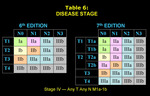
DISEASE STAGE
The combinations of T,
N,
and M grades that constitute a disease stage were changed in the 7th edition of the TNM staging system as a result of the revisions to the T and M descriptors described above.
The disease stages will be defined under the 7th edition of the TNM staging system as exposed in Table 6 (which includes the 6th edition for comparison).
The major changes include the following:
- Reclassification of T2bN0M0 as stage IIA,
instead of stage IB.
- Reclassification of T2aN1M0 as stage IIA,
instead of stage IIB.
- Reclassification of T3 (>7cm)N0M0 as IIB,
instead of stage IB.
- Reclassification of T3 (>7cm)N1M0 as IIIA,
instead of stage IIB.
- Reclassification of T3N0M0 (separate tumor nodules in the same lobe as the primary tumor) as stage IIB instead of stage IIIB.
- Reclassification of T3N1M0 or T3N2M0 (separate tumor nodules in the same lobe) as stage IIIA instead of stage IIIB.
- Reclassification of T4N0-1M0 as stage IIIA,
instead of IIIB
- Reclassification of T4N2-3M0 as stage IIIB,
instead of stage IV.
- Reclassification of malignant pleural effusion (M1a) as stage IV,
instead of stage IIIB.
Survival: The median survival correlates with both the clinical stage and surgical-pathologic stage under the 7th edition of the TNM staging system:
- Clinical stages IA,
IB,
IIA,
IIB,
IIIA,
IIIB,
and IV have a median survival of 60,
43,
34,
18,
14,
10,
and 6 months,
respectively.
- Surgical-pathologic stages IA,
IB,
IIA,
IIB,
IIIA,
IIIB,
and IV have a median survival of 119,
81,
49,
31,
22,
13,
and 17 months,
respectively.
The patients with stage IV disease have unusually good survival because the estimate is based on a small number of patients who were well enough to undergo surgical resection.

")
")
")
 The IASLC lung cancer staging project: a proposal for a new international lymph node map in the forthcoming seventh edition of the TNM classification for lung cancer. J Thorac Oncol 5:568-577")

. Stage T1a tumors are those ≤2 cm.")
.")
.")

. Note the presence of a separated nodule in the same lobe as the primary tumor. Findings are consistent with a stage T3 tumor.")
 presenting with massive mediastinal invasion (trachea, carina, aortic arch, esophagus). Right pulmonary artery encasement, abrupt amputation of the right upper lobe and narrowing of the right main-stem brinchus are observed. Parietal pleural invasion in the RUL and pleural effusion (solid arrowhead) are also depicted. These findings are consistent with a stage T4 tumor. Contralateral hilar adenopathies (solid arrow) are shown, indicating N3 lymph nodes. Note the presence of separate tumor nodules in the right middle lobe (open arrow) and left lower lobe (open arrowhead). So far, these findings are indicative of a M1a tumor. The presence of a right costal lytic lesion (green circle) with associated soft tissue mass, multiple hepatic hypodense lesions (blue circles) and a right adrenal mass are consistent with a stage T4N3 M1b tumor.")