First,
the BNU index were measured on both type of imager. The BNU index was respectively 11 % and 16% for the 9" and 12" XRIIs.
For the XFPD,
the BNU index was 0,6 % in both binnings 1x1 (pixel size = 0,184 mm) and 2x2 (pixel size = 0,368 mm).
Therefore,
the XFPD shows better uniformity than XRIIs.
The step wedge measures were performed at 4 different doses level (9-18-40-75 nGy) at 70 kVp.
These measures between the 3 image sensors (figure 3) shows that the XFPD has a better linear response than a RIU image sensor.
Moreover,
the RIUs signals saturate at 40 nGy as opposed to the XFPD which has a still a linear behaviour at 75 nGy
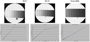
Fig.: 3 : step wedges and linearity measures
The CNR was measured on the 3 image sensors in the same dose conditions (50 kVP and 70 nGy per frame) and for each sensor 16 images were averaged. The CNR was respectively 14,
12 and 11 for the XFPD (binning 1x1) 9¨XRII and 12"XRII. Therefore,
the XFPD offers a slightly better CNR than conventional XRII.
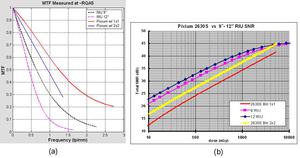
Fig.: 4 : MTF (a) and SNR (b) measures at RQA 5 conditions
The MTF and SNR measures for the 3 image sensors were performed in the RQA5 and RQ9 conditions (figure 4).
MTF curves shows that the XFPD has significant better spatial resolution.
For instance the XFPD has an MTF(1lp/mm) = 0,48 compared to MTF(1lp/mm) = 0,1 for the 12¨RII.
On the other hand,
the SNR is better for the XRII 12¨than the XPD in binning 12 at low dose performances.
Indeed,
the SNR at 100 nGy is 33 dB for the 12¨XRII and 28 dB for the XFPD (binning 2x2).
Then,
two sets of pelvis acquisitions were performed on the 3 image sensors.
For the XFPD,
image acquisitions were performed with two different binnings (1x1 and 2x2).
The first set acquisitions was performed at 12 nGy per frame (80 kvP,
0,6 mA) with the antiscattering grid.
The second set of acquisitions was performed adding a 10 cm lucite bloc on the pelvis phantom at 25 nGy per frame (116 kvP,
1,8 mA) without the antiscattering.
Each set of acquisition (ie: 10 images) were averaged and then displayed on a monochrome monitor.
The image quality for the 3 sensors at 12 nGy per frame (figure 4) shows that 9-12" XRII's and the XFPD (binning 2x2) has comparable behaviour (figure 5).
Indeed,
the bone details (shape and edges) have the same visibility in both type of images (XRII's and XFPD) On the other hand,
the XFPD binning 1x1,
the bone visibility is poor due to a higher noise.
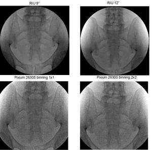
Fig.: 5 : 80kV and 12 nGy/fr detector entrance dose
As the noise is higer in a XFPD binning 1x1 (lower SNR) and as both 9-12 RIUS have similar behaviours,
a second visual comparison was performed between a 12" RIU and a XFPD binning 2x2 at 25 nGy entrance and with a 10 cm lucite thickness (eq ~30 cm patient thickness).
Both image sensors show similar image quality at low dose performances (figure 6).
Bone visibility (shape and edges) is comparable in both type of images.
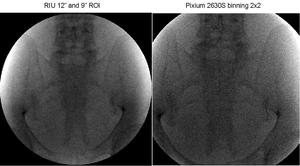
Fig.: 6 : Heavy patient anatomy (pelvis + 10 cm lucite), 116kV, 25 nGy/fr,
In order to have an objective visual assessment between two image sensors having different spatial resolutions (ie: MTF), the ratio of the XFPD in binning 1x1 (resp 2x2) MTF to the 12"RIU has been applied on the XFPD images acquired at the 12 nGy per frame entrance dose. This processing was applied in the fourier domain with the same sampling factor (c.f.
figure 7).
In this context,
the XFPD acquired in a binning 1x1 has a better bone visibility which is nearly similar to the 12"RIU images.
Moreover,
the XFPD acquired in a binning 2x2 has a better image quality (visibility and sharpness) than the 12"RIU data after the MTF compensation processing.
Thus,
both imagers reach equivalent image quality performances at a very low entrance dose.
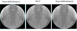
Fig.: 7: XFPD/12"RIU MTF ratio processing and resampled with identical sampling factor (300x300 image size)
Aknowledgement : The authors would like to thank Karine Bigot Moirans/FR for the image data analysis performed on the XRII and XFPD imagers.
