ECR 2012 / C-0499
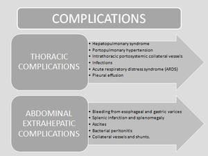
Thoracic and abdominal extrahepatic complications of liver cirrhosis : what the radiologist should know?
Congress:
ECR 2012
Poster Number:
C-0499
Type:
Educational Exhibit
Keywords:
Cirrhosis, Complications, CT, Liver
Authors:
E. Canales Lachén, B. Diaz-Barroso, D. Mollinedo, M. C. Pulido Rozo, S. Martín Pérez, C. Rubio Hervás, A. Teruel; Madrid/ES
DOI:
10.1594/ecr2012/C-0499

Fig. 1

Fig. 9:
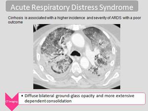
Contrast-enhanced CT scan shows diffuse ground-glass opacity in both lungs and...

Fig. 14

Fig. 15

Fig. 16

Fig. 17

Fig. 18

Fig. 19

Fig. 20

Fig. 21

Fig. 13

Fig. 10

Fig. 11

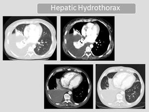
Fig. 8:
Contrast –enhanced CT scans show a large amount of pleural effusion in the...

Fig. 7

Fig. 5

Fig. 6:
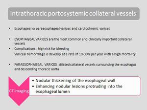
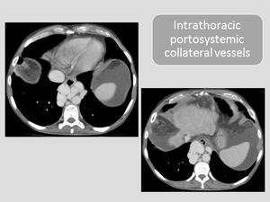
Axial contrast-enhanced CT images show dilated paraesophageal varices and...

Fig. 12

Fig. 22
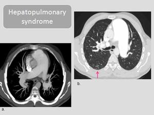
 Axial thin-slab maximun intensity projection CT image shows enlarged pulmonary arteries and slightly dilated subpleural vessels.
(b)CT scan in lung window shows the dilated subpleural vessels that extend toward the lung periphery and nodular dilatation of peripheral pulmonary vessels (arrow).
(Courtesy of Dra.Torres and Dra.Fernández-Velilla. Hospital La Paz, Madrid, Spain)
References: Dra. Torres and Dra. Fernández-Velilla. Department of Radiology, Hospital La Paz, Madrid, Spain")
Fig. 4:
Hepatopulmonary syndrome. (a) Axial thin-slab maximun intensity projection CT...
 References: Dra. Torres and Dra.Fernández-Velilla. Department of Radiology.Hospital La Paz, Madrid, Spain")
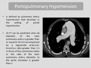
Fig. 2:
Contrast-enhanced CT scan shows a widened main pulmonary artery with a...

Fig. 3