Type:
Educational Exhibit
Keywords:
Parasites, Infection, Cysts, Education, Diagnostic procedure, Complications, Ultrasound, CT, Conventional radiography, Biliary Tract / Gallbladder, Abdomen, Liver
Authors:
A. Batista1, M. Baptista1, M. A. A. Vieira1, V. B. Monteiro2, E. M. G. Batista3, M. A. Alves1, M. Matias1; 1Beja/PT, 2Loures/PT, 3Lisbon/PT
DOI:
10.1594/ecr2012/C-2270
Imaging findings OR Procedure details
The authors illustrate hydatid disease as a dynamic entity with multiple imaging appearances and different parasitizing locations,
reviewing 27 patients evaluated in our institution with hepatic (24),
pulmonary (1),
gastric tract (1) and soft tissue (1) hydatid cysts,
as well as demonstrating radiological appearances of hydatid cyst complications in the form of rupture (4),
infection (1) and common bile duct compression (1),
with cases of representative patients.
The radiological diagnosis were confirmed by reviewing clinical and surgical records.
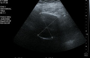
Fig. 1: Ultrasonography demonstrating a type CE5 hepatic hydatid cyst, with visible internal echoes and a thick calcified wall.
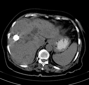
Fig. 2: Multidetector CT transversal image showing a totally calcified hydatid hepatic cyst in segment V.
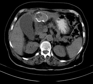
Fig. 3: Low density oval mass with partially calcified wall and interruption of contour representing ruptured left lobe hepatic hydatid cyst comunicating with intrahepatic biliary ducts.
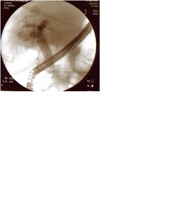
Fig. 4: ERCP of hydatid cyst pictured in the previous CT image demonstrating bile duct-cyst wide communication after hydatid cyst rupture.
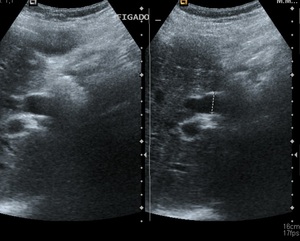
Fig. 5: Marked common bile duct dilation due to bile duct-cyst wide communication after hydatid cyst rupture (demonstrated by posterior ERCP).
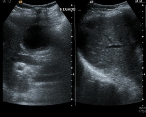
Fig. 6: Gallblader and intrahepatic bile duct dilation due to bile duct-cyst wide communication after hydatid cyst rupture (demonstrated by posterior ERCP).
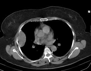
Fig. 7: Pulmonary hydatid cyst extending to the lateral chest wall.
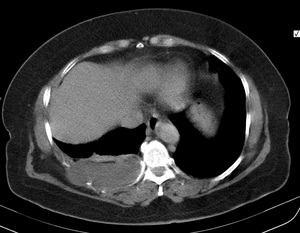
Fig. 8: Pulmonary hydatid cyst extending to the posterior chest wall.
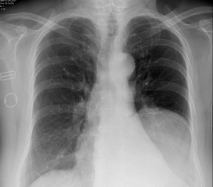
Fig. 9: Postero-anterior thoracic radiogram with well defined nodular hyperlucency in the the lower left lobe basal segments corresponding to a pulmonary hydatid cyst.
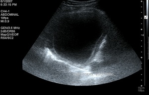
Fig. 10: Ultrasound caracterization of previous radiogram showing a type CE1 cyst.
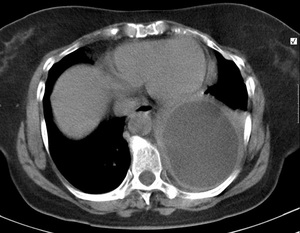
Fig. 11: Low density content pulmonary hydatid cyst
demonstrated by multidetector non-enhanced CT.
