ECR 2012 / C-2495
Intraabdominal Active Bleeding: Helical CT , MDCT (64-slice), DSA and Homeostatic Embolization Findings
Congress:
ECR 2012
Poster Number:
C-2495
Type:
Educational Exhibit
Keywords:
Haemorrhage, Embolisation, CT-Angiography, CT, Catheter arteriography, Emergency, Abdomen
Authors:
B. ALPARSLAN1, N. YILDIRIM1, B. Hakyemez2, A. Ozer1, G. Savci1; 1Bursa/TR, 2Türkiye/Bursa/TR
DOI:
10.1594/ecr2012/C-2495

Fig. 1
At the hilum of the liver, just anterior to the portal vein, high density contrast extravasation with irregular margins(↓) and surrounding hematoma(→) can be seen.
Helical CT, delayed phase (5-10’) (b)Contrast media leaks from the injured artery and spreads through the perihepatic space (←).")
Fig. 2:
Case 1: 23, M, trauma, hepatic artery injury
Helical CT, 60’’ image (a)At...
 and severity of bleeding, MDCT and MDCT-A was performed. Extravasation within hematoma (↓) is clearly seen in arterial phase (a) and extravasation becomes more prominent (←) in the portal phase (b). (also seen splenic injury and perisplenic hematoma)")
Fig. 3:
To get further information about which vessel was effected (artery/venula) and ...
 from hepatic artery (just distal to the gastroduodenal artery bifurcation) was determined (a).
With selective catheterization the extravasation became more prominent (→)(b).And the contrast agent spread to the perihepatic area (↑, c).")
Fig. 4:
DSA was performed 30 min after MDCT and extravasation (↓) from hepatic artery...
. The patient was hospitalized for other traumatic injuries and was discharged from the hospital after full recovery.")
Fig. 5:
The bleeding was stopped by urgent coil embolization (↓). The patient was...
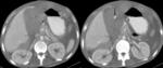
Fig. 6:
Case 2: 26, M, trauma, hepatic artery injury
Helical CT, 60’’ image: In...
 and hematoma at the pancreatic tail region; laceration at the inferior pole of spleen(*) were detected.")
Fig. 7:
Case 3: 43, M, trauma, spleen
Helical CT, 60’’ image: Active contrast...
 lack of active contrast extravasation was in favour of bleeding from venous origin. Venous phase coronal reformatted images (b,c) showed enlarging hematoma(*) related to the inferior pole of spleen (←).
The patient was discharged uneventfully after conservative therapy.")
Fig. 8:
Multiphasic MDCT was performed 2h later: In arterial phase (a) lack of active...
, trauma, spleen
Helical CT, unenhanced(a) & 60’’ image (b)Hyperdense, spheric hematoma is clearly seen in unenhanced image (a, *).
In portal venous phase image (b) contrast extravasation area isodense to vascular structures around (+) is depicted. Patient underwent splenectomy and bleeding was controlled but was lost due to other reasons.")
Fig. 9:
Case 4: 51, M, Hodgkin Lymphoma (+), trauma, spleen
Helical CT, unenhanced(a)...
 gastric wall irregularity is also seen(←) in addition to perihepatic fluid.In venous phase image (c) contrast blush (isodense to aorta) is seen within gastrohepatic ligament (↓).
Patient underwent surgery, in laparotomy gastric perforation and gastric artery injury were detected and successfully repaired.")
Fig. 10:
Case 5: 38, M, trauma, gastric perforation
Helical CT (NECT and 60’’...
: Hyperdense active bleeding spot is seen within the right lobe of liver (a,↓) but due to the lack of multiphasic imaging it is hard to differentiate arterial-portal venous or hepatic venous origin. Three days later control CT was performed (b). The patient was treated conservatively and recovered uneventfully.")
Fig. 11:
Case 6: 47, M, trauma, liver hematoma
Helical CT (60’’ images):...
, coronal (b) and sagittal (c) reformatted images: In some patients active bleeding(↓) can be present irrelevant with the prediagnosis (in this case intraabdominal mass was suspected). Radiologists should be aware of the findings and immmediately inform the clinician. The patient was hemodynamically stable in CT suit but became unstable in his room and was lost despite vigorous resuscitation.")
Fig. 12:
Case 7: 63, M, multiple myeloma
MDCT 70’’ axial image(a), coronal (b) and...
: Images of pelvic (a), liver (b) and umblicus (c) levels demonstrate high density, intraperitoneal fluid (a,↓), mesenteric haziness on the right side (c,↑)and also thickening of left anterior renal fascia (c, →). The patient was treated conservatively and recovered uneventfully.")
Fig. 13:
Case 8: 79, F, trauma
Helical CT (60’’): Images of pelvic (a), liver (b)...
: Images of pelvic (a), liver (b) and umblicus (c) levels demonstrate intraperitoneal fluid with increased density (a,↓). The fluid also fills the perihepatic area (b,→). Mesenteric haziness on the right side became more prominent (c,↑). Findings favour active bleeding. The patient was treated conservatively and recovered uneventfully.")
Fig. 14:
Helical CT (15`): Images of pelvic (a), liver (b) and umblicus (c) levels...
 and was controlled after coil embolization.")
Fig. 15:
Case 9: 82, M, massive rectal bleeding, angiodysplasia
DSA images of the...

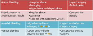
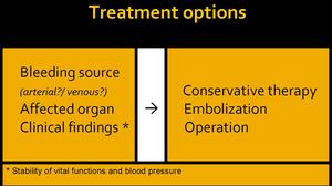
Table 3