ECR 2013 / C-1216
Radiological assessment of skull sutures in Patients with syndromic craniosynostosis.
Congress:
ECR 2013
Poster Number:
C-1216
Type:
Educational Exhibit
Keywords:
Anatomy, Neuroradiology brain, Paediatric, CT-High Resolution, MR, CT, Computer Applications-3D, Computer Applications-Detection, diagnosis, Computer Applications-General, Cerebrospinal fluid, Congenital, Image verification
Authors:
G. D'Apolito, R. Calandrelli, G. Di Lella, A. M. Costantini, C. Colosimo; Rome/IT
DOI:
10.1594/ecr2013/C-1216

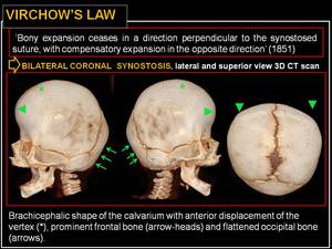
Fig. 2:
Virchow’s law.
: sutures involved. References: Department of Bio-imaging and Radiological Sciences, Catholic University of Rome - Rome/IT")
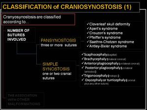
Fig. 3:
Classification of craniosynostosis (1): sutures involved.
: association with other malformations. References: Department of Bio-imaging and Radiological Sciences, Catholic University of Rome - Rome/IT")
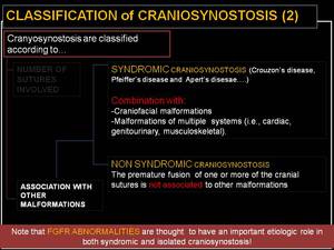
Fig. 4:
Classification of craniosynostosis (2): association with other malformations.
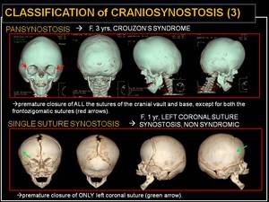
: pansynostosis VS single suture synostosis. References: Department of Bio-imaging and Radiological Sciences, Catholic University of Rome - Rome/IT")
Fig. 5:
Classification of craniosynostosis (3): pansynostosis VS single suture...

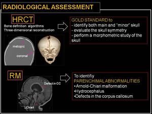
Fig. 6:
Radiological assessment.
: the different arches drawn on the skull. References: Department of Bio-imaging and Radiological Sciences, Catholic University of Rome - Rome/IT")
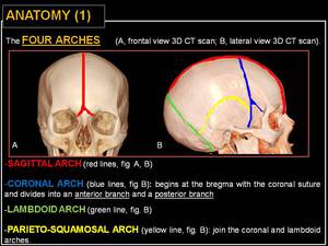
Fig. 7:
Anatomy (1): the different arches drawn on the skull.
: sagittal arch. References: Department of Bio-imaging and Radiological Sciences, Catholic University of Rome - Rome/IT")
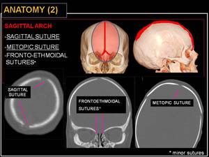
Fig. 8:
Anatomy (2): sagittal arch.
: coronal arch. References: Department of Bio-imaging and Radiological Sciences, Catholic University of Rome - Rome/IT")
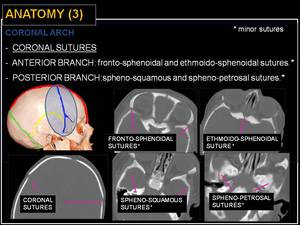
Fig. 9:
Anatomy (3): coronal arch.
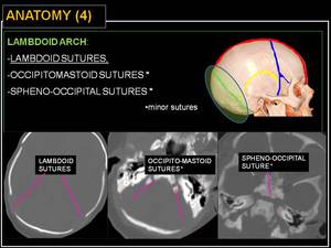
: lambdoid arch. References: Department of Bio-imaging and Radiological Sciences, Catholic University of Rome - Rome/IT")
Fig. 10:
Anatomy (4): lambdoid arch.
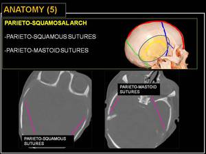
: parieto-squamosal arch. References: Department of Bio-imaging and Radiological Sciences, Catholic University of Rome - Rome/IT")
Fig. 11:
Anatomy (5): parieto-squamosal arch.