ECR 2013 / C-2018
Usefulness of lung biopsy with CT-guided coaxial technique in histological diagnosis of lung cancer
This poster was previously presented in Spanish at the 2012 Congreso Nacional SERAM (Granada)
Congress:
ECR 2013
Poster Number:
C-2018
Type:
Scientific Exhibit
Keywords:
Lung, Oncology, Interventional non-vascular, CT, Percutaneous, Biopsy, Neoplasia
Authors:
J. A. Gallego Sánchez, N. Riera Bevia, B. Pomares Rey, Z. Sánchez Acevedo, E. Ramos Gavila, E. Alsina Seguí; Elche/ES
DOI:
10.1594/ecr2013/C-2018

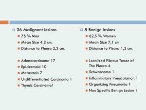
Table 1:
Characteristics of lesions

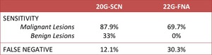
Table 2:
Sensitivity and false negative rate in samples obtained with 20G-SCN and 22G-FNA

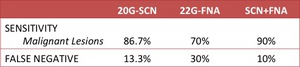
Table 3:
Sensitivity and false negative rate in samples obtained with 20G-SCN and...