
ECR 2014 / C-0959
Cerebellar tonsil herniation: Its diverse pathogenesis
This poster is published under an open license. Please read the disclaimer for further details.
Congress:
ECR 2014
Poster Number:
C-0959
Type:
Educational Exhibit
Keywords:
CNS, Anatomy, MR, Diagnostic procedure, Cerebrospinal fluid, Congenital, Hernia
Authors:
H. Mukai, H. Yokota, T. Horikoshi, K. Motoori, T. Uno; Chiba/JP
DOI:
10.1594/ecr2014/C-0959

Fig. 5:
Chiari malformation

Fig. 6:
Crouzon disease
Shallow posterior fossa and CTH are seen on CT

Fig. 7:
Patient suspected Pfeiffer syndrome
Radiohumeral synostosis is seen on...

Fig. 8:
Patient suspected Pfeiffer syndrome
Craniosynostosis involving the lambdoid...

Fig. 9:
Intracranial space-occupying lesions
Not only infratentorial lesions but also...

Fig. 10:
Acromegaly

Fig. 11:
Spontaneous intracranial hypotension
Thick dural enhancement, bilateral thin...

Fig. 12:
Hydrocephalus caused by aqueduct stenosis
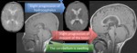
Fig. 13:
Macrocephaly capillary malfomation
(Macrocephaly-cutis marmorata...