The Equipment:
All examinations were carried out using a multi-frequency linear array probe (6 - 10 MHz,
Shimadzu Co.
Ltd.).
The Technique of Examination:
The patients were positioned supine with neck extended.
Examination was done through closed eyelids.
The 10 MHz linear array probe was applied over the eyelid,
after applying sufficient quantity of ultrasound gel.
This helped in achieving better contact between the probe and the eyelid.
The conscious patients were directed to keep their eyes in neutral (mid) position and to suppress eye movement.
The probe was kept in such a position so as to achieve axial images of optic nerve.
This was confirmed by typical appearance of optic nerve seen in this section.
(See figure 1).
We chose to measure optic nerve diameter in axial images based on the evidence provided by experimental study of Hansen and Helmke in which in vivo optimum accuracy of optic nerve diameter measurement was seen to be achieved by axial sections.
Once the required section was achieved,
the gain settings were reduced to minimum to achieve optimum image quality.
Optic nerve diameter was measured at a distance of 3 mm from optic nerve head (FIGURE 1)
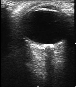
Fig. 1: AXIAL IMAGE OF ON, SHOWING TYPICAL APPEARANCE OF ON.
References: Department of Diagnostic radiology, King George Medical College, Lucknow, U.P, India.
FIGURE 1: AXIAL IMAGE OF ON,
SHOWING TYPICAL APPEARANCE OF ON.
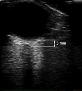
Fig. 2: ON IS BEING MEASURED AT 3 MM POSITION (THE BULBOUS PART OF ON).
References: Department of Diagnostic radiology, King George Medical College, Lucknow, U.P, India.
FIGURE 2: ON IS BEING MEASURED AT 3 MM POSITION (THE BULBOUS PART OF ON).
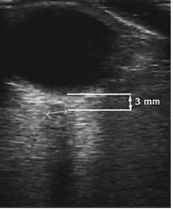
Fig. 3: THE OND WAS MEASURED AT 3 MM POSITION IN A DIRECTION PERPENDICULAR TO AXIS OF ON.
References: Department of Diagnostic Radiology, King George Medical College, Lucknow, U.P, India.
FGURE 3: THE OND WAS MEASURED AT 3 MM POSITION IN A DIRECTION PERPENDICULAR TO AXIS OF ON.
The patients & Selection protocol:
Patients were selected from those admitted in department of medicine and pediatrics,
KGMC (CSSMU).,
Lucknow,Uttar Pradesh,
India.
Patients were also selected from those attending our (Radiology) department for ultrasound examination.
All patients greater than one year age were eligible for inclusion into study.
Patients less than one year of age were excluded from the study.
The patients were divided into following four groups:–
Group I (Controlled) (n=27 + 9 = 36): Definition: Patients attending our department for ultrasound examination for reasons not related to any condition that may lead to raised intra cranial pressure or change in optic nerve diameter.
Group II (n=15) Definition: Patients with jaundice or portal hypertension or with history of chronic liver disease,
but with no signs of hepatic encephalopathy
Group III (cases) (n=22): Definition: Patients diagnosed as hepatic encephalopathy.
Group IV (n=5): Definition: Patients with stroke (proven by CT scan)/ encephalopathy due to cause other them hepatic encephalopathy.
Follow-up:
Cases were followed for duration of 30 days.
At the end of 30 days follow up the outcome was labeled as either death/ did not improve or survival/ improved.
Statistical analysis:
All data was entered in a Microsoft Office,
Excel spread sheet.
The average optic nerve diameter of both eyes was derived from the 4 readings taken for each optic nerve at the 3 mm point (this was labeled as “averaged optic nerve diameter” - AOND)
The final optic nerve diameter of patients was calculated by averaging the AOND of each eye,
and labeled as “optic nerve sheath diameter” – ONSD.
The average optic nerve diameter for a given group was calculated as an average of ONSD of each individual in that group and labeled as mean optic nerve sheath diameter (MONSD) of that group.
Criterion used to diagnose a given optic nerve as dilated:
In the present study no predetermined parameter was used to classify optic nerve as dilated or non-dilated.
Any optic nerve diameter reading that was greater than MONSD+2 SD of controls greater than 5 years was considered to be outside normal range.


. References: Department of Diagnostic radiology, King George Medical College, Lucknow, U.P, India.")
