ECR 2014 / C-1997
Data of sonography allow to model of malignancies growth in infants
This poster is published under an open license. Please read the disclaimer for further details.
Congress:
ECR 2014
Poster Number:
C-1997
Type:
Scientific Exhibit
Keywords:
Abdomen, Oncology, Paediatric, Ultrasound, Ultrasound-Colour Doppler, Ultrasound-Power Doppler, Diagnostic procedure, Staging, Screening, Cancer, Congenital, Pathology
Authors:
I. Begun, O. Krasko; Minsk/BY
DOI:
10.1594/ecr2014/C-1997
")
Fig. 9:
Nonlinear model using the Gompertz function describing the growth of the tumor...

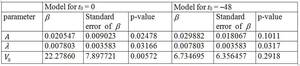
Fig. 10:
Table 1
")
Fig. 11:
Estimates of the tumors volumes depending on infants ages with the...

Fig. 12:
Table 2