This poster is published under an
open license. Please read the
disclaimer for further details.
Keywords:
Breast, Oncology, MR-Diffusion/Perfusion, MR, CAD, Computer Applications-Detection, diagnosis, Technology assessment, Tissue characterisation
Authors:
H. Dijkstra, M. D. Dorrius, M. Wielema, M. Oudkerk, P. E. Sijens; Groningen/NL
DOI:
10.1594/ecr2015/B-0624
Methods and materials
Twenty-eight consecutive patients with breast lesions ≥ 1 cm were examined with 1.5T DWI (b=0,50,200,500,800,1000 s/mm2) between July 2012 and June 2013.
Lesions were classified by histopathology or follow-up and non-mass lesions were excluded.
Offline IVIM voxel-by-voxel analysis (Matlab,
The Mathworks,
Natick,
MA,
USA) yielded molecular diffusion (Dslow),
microperfusion (Dfast) and respective fractions (fslow/fast) using equation 1 (Fig.
1).
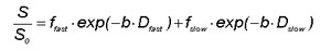
Fig. 1: IVIM equation
For each lesion,
voxels were selected by drawing a large ROI around each lesion on the b = 0 s/mm2 DWI image (Fig.
2).

Fig. 2: Example of lesion ROI
Then,
Dslow,
Dfast and ffast were defined for each lesion by the median of the first X percentage of the highest values (top X%) within the ROI,
where X varied between 1% (highest pixel values of the ROI) and 100% (all pixels of the ROI) in steps of 1%; this procedure yielded 100 datasets for each lesion.
Subsequently,
for each of the 100 datasets,
the three IVIM parameters were combined in parallel in a single test using the “AND rule” to exclude malignancy,
based on three thresholds [4].
That is,
if Dslow is lower than the threshold,
and if Dfast is lower than the threshold,
and if ffast is lower than the threshold,
then the combined result is defined positive (malignant),
but in all other cases,
the combined result is negative (benign).
The dataset which yielded the highest number of true negatives was used as the final result,
and the respective thresholds and topX% were recorded.
