This poster is published under an
open license. Please read the
disclaimer for further details.
Keywords:
Thyroid / Parathyroids, Ear / Nose / Throat, Oncology, Ultrasound, Diagnostic procedure, Biopsy, Tissue characterisation, Pathology
Authors:
B. Raghavan, D. Sundaram; Chennai/IN
DOI:
10.1594/ecr2015/B-0877
Methods and materials
Inclusion criteria:
- Patients with thyroid swelling or palpable thyroid nodule.
- Patients who were found to have thyroid nodules incidentally during CT scan of neck or chest for some other purpose.
Exclusion Criteria:
- Maximum nodule diameter < 5 mm.
- Completely anechoic lesion (cyst) with no solid component,
which did not require fine needle aspiration cytology.
- Inconclusive cytology due to inadequate sample or hemorrhagic material.
Sample size:
86 patients with 141 nodules were studied,
out of them 79 patients with 130 nodules were included in the study.
(Fig. 3)
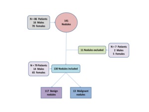
Fig. 3: Flow chart of selection of patients.
PROCEDURE:
All the nodules were evaluated with ultrasound as well as VTQ and VTI methods of ARFI elastography technique and grouped according to TIRADS category based on ultrasound findings.Normal thyroid parenchyma (category1) and biopsy proven malignant nodules (category 6) were excluded.
In VTQ method,
5 measurements were taken for each nodule and the average was considered as the VTQ value for that nodule.
Areas of calcification,
cystic and necrotic components were not included in the ROI box.
The shear wave velocity range for the machine used in the study was 0.5 – 8.4 m/s.
Velocity greater than this was expressed as x.xx m/s.
When x.xx m/s was seen in a solid nodule it was recorded as the maximum shear wave velocity,
i.e.
8.4 m/s (6)(7)(8) .
In VTI method,
nodules appear brighter than adjacent parenchyma considered as soft ,
darker than adjcacent parenchyma considered as hard and equal to the adjcacent parenchyma as isoelastic (9).
Under ultrasound guidance,
aspiration was done using 23 G needle attached to a 10 ml disposable syringe with to and fro movement of the needle and a gentle suction with the syringe.
Statistical Analysis:
Data validation and analysis was carried out by SPSS (Statistical Package for Social Sciences) version 11.0 and P value <0.05 was considered as statistically significant.The imaging findings were correlated with fine needle aspiration cytology results.
