1.WRIST
Anatomy
It is known that the carpal bones are divided into two rows: proximal and distal. The proximal carpal row is represented by the scaphoid,
lunate,
triquetrum,
and pisiform while the distal carpal row is comprised of the trapezium,
trapezoid,
capitate,
and hamate.
The distance between carpal bones is less than 3 mm.
Three smooth arches along carpal: a first arch that is a smooth curve outlining the proximal convexities of the scaphoid,
lunate and triquetrum,
a second arch that traces the distal concave surfaces of the same bones,
and the third arch which follows the main proximal curvatures of the capitate and hamate.
(Fig.1)

Fig. 1: Plain radiography of the left wrist - posteroanterior view - X-ray anatomy of carpal bones.
References: SCJU - UPU / Cluj-Napoca /RO
SCAPHOID FRACTURE
The scaphoid is one of the most fractured carpal bone that occurs in young and middle-aged adults.
About 10% of the scaphoid fractures have associated fractures of other bones.The most frequently interest zones are: the radial styloid,
but also triquetrum,
capitates and perilunate fracture-dislocations.
Approximately 1% of scaphoid fractures are bilateral.
Mechanism:
The mechanism of the injury is usually a fall on an outstretched hand (F.O.O.S.H.),
with a dorsiflexion of the hand,
but it also may include a direct impact.
Exam: The patients present in the emergency room with a specific symptomatology as is the tenderness of the "anatomic snuffbox",
pain at percussion over the scaphoid tubercle and pain in the snuffbox with the ulnar pronation of the wrist.
X Ray: Sometimes the initially radiographs of a scaphoid fracture may appear normal.
Patients with history of a fall on the outstretched hand that present with a pain in the snuffbox,
should be orthopedically immobilized and sent for additional imaging studies.
The radiologist technician should consider obtaining additional scaphoid views like a posteroanterior view of the wrist,
with the wrist positioned in 45 degrees of ulnar deviation and pronated obliquely at 45 degrees.
If clinically suspected,
a scaphoid fracture should be treated as such even if the standard series of X-rays shows no fracture.
Patients wrist should be orthopedically immobilized and followed up clinically in 10 days with repeat X-rays as required.
The importance of the scaphoid fracture is due to the complications that can appear if the fracture is missed on X ray and if the patient does not receive immediately orthopedically treatment.
The most frequent complications are avascular necrosis and the nonunion of the fracture.
(Fig.2; Fig.3; Fig.4)
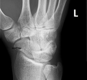
Fig. 2: Plain radiography of the left wrist - posteroanterior view - incomplete fracture of the scaphoid in a male pacient - 30 years old.
References: SCJU - UPU / Cluj-Napoca /RO
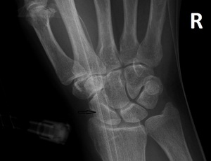
Fig. 3: Plain radiography of the right wrist - posteroanterior view - incomplete fracture of the scaphoid in a female pacient - 61 years old.
References: SCJU - UPU / Cluj-Napoca /RO
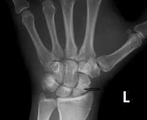
Fig. 4: Plain radiography of the left wrist - posteroanterior view - fracture of the scaphoid in a male pacient - 22 years old.
References: SCJU - UPU / Cluj-Napoca /RO
2.ELBOW
Anatomy
The elbow joint is made up of three articulations :
- Radiohumeral:capitellum of the humerus with the radial head
- Ulnohumeral:trochlea of the humerus with the trochlear notch of the ulna
- Radioulnar: radial head with the radial notch of the ulna.
(Fig.5)
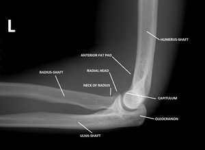
Fig. 5: Lateral view of the left elbow - X-ray anatomy with normal fat pad.
References: SCJU - UPU / Cluj-Napoca /RO
RADIAL HEAD FRACTURE
The radial head fractures are relatively common injuries,
occurring in about 20% of all acute elbow injuries,
especially in adults.
Fractures of the radial head can be occult on radiographs.
Mechanism:
The mechanism of injury is usually a result of indirect trauma like a fall on an outstretched arm.
Although,it could be a direct blow to the elbow that can cause a radial head fracture,
but this is uncommon.
Exam:
The patient presents in the emergency room with a swelling over the lateral side of the elbow and with limited range of motion: heavily forearm rotation and elbow extension.
Xray:
Standard radiographic evaluation of radial head fractures includes AP and lateral views of the elbow,
although an oblique view is very frequently also obtained for better visualize the radial head.
On the elbow x rays,
the fat pads can be seen.
They are collections of fat tissue adjacent to elbow joint capsule that appears as lucency on x rays.
The anterior fat pad can be normal but if it is displaced and elevated,
is pathologic and it is named “sail sign”.
If a posterior fat pad can be seen,
that is abnormal and is an indirect sign of fracture.
Often a non-displaced radial head fractures can be easily missed on plain films if there is no attention given to the indirect signs. (Fig.6; Fig.7).
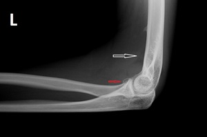
Fig. 6: Lateral view of the left elbow - subtle radial head fracture with sail sign present in a young female - 20 years old.
References: SCJU - UPU / Cluj-Napoca /RO
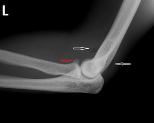
Fig. 7: Lateral view of the left elbow - subtle radial head fracture with anterior sail sign and posterior fat pad in a young male - 23 years old.
References: SCJU - UPU / Cluj-Napoca /RO
3.FOOT
Anatomy
The bones of the foot are divided into three categories:
- tarsal
- metatarsal
- phalanges.
The tarsal bones of the foot are organised into three rows:
- proximal: the talus and the calcaneus
- intermediate: the navicular
- distal: the cuboid and the three cuneiforms
The forefoot contains the metatarsals and the phalanges.
(Fig.8)
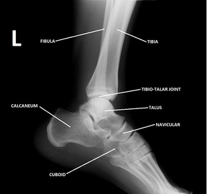
Fig. 8: Lateral view of the left foot - X-ray anatomy.
References: SCJU - UPU / Cluj-Napoca /RO
CALCANEUS FRACTURE
The calcaneus is the most commonly fractured tarsal bone and accounts for about 2% of all fractures and ~60% of all tarsal fractures.
Mechanism:
Calcaneus fracture often result from falling from height but may be due to more trivial injury.
Exam:
The most common symptoms of a calcaneus fracture are: pain,
bruising,
swelling,
heel deformity and inability to put weight on the heel or walk.
Xray:
Lateral X-rays of the calcaneus show Bohler's angle.
Bohler's angle is the angle between two tangent lines drawn across the anterior and posterior borders of calcaneus in the lateral view.
When Bohler's angle becomes less than 20 degrees it indicates a calcaneal fracture.
The Bohler’s angle is an angle seen on the lateral view of the foot.
It is the angle between a line bordering the superior aspect of the posterior calcaneal tuberosity and the superior subtalar articular surface and a line crossing through the superior subtalar articular surface and the superior aspect of the anterior calcaneal process.
The Bohler’s angle is normally 20-40 degrees.(Fig.9; Fig.10)
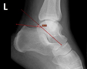
Fig. 9: Lateral view of the left foot - normal Bohler's angle.
References: SCJU - UPU / Cluj-Napoca /RO
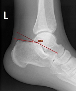
Fig. 10: Lateral view of left foot - Bohler's angle - less than 20 degrees.
References: SCJU - UPU / Cluj-Napoca /RO


