Subjects
We prospectively enrolled 44 consecutive diagnostic breast MRI scans.
DKI was applied as a part of our standard breast MRΙ protocol in combination with dynamic contrast enhanced (DCE) -MRΙ in all cases.
The referral indications were mostly two: preoperative local staging of the tumor and clarification of suspicious findings on conventional imaging (mammography,
ultrasound) or clinical examination.
Imaging Protocol
All patients were examined in a 1.5 Tesla (T) MR unit equipped with a four-channel dedicated double breast coil.
Consensus of the examined women was initially taken .Whenever appropriate,
we performed the scan during the second week of the menstrual cycle for the premenopausal women.
Patients were placed in prone position.
The conventional imaging protocol included an axial fat suppressed T2-weighted turbo spin-echo sequence (repetition time/echo time [TR/TE]: 4000/97; matrix: 320×320 pixels; slice thickness: 3 mm); a T1-weighted non-fat suppressed sequence; and a T1-weighted fat suppressed DCE sequence with one pre-contrast and six post-contrast acquisitions in the axial plane (dynamic scan duration: 1 min,
slice thickness: 1.5 mm).
An MIP projection was also taken.
DKI was obtained before dynamic images.
Two-dimensional (2D) spin-echo echo-planar imaging (EPI) sequence (TR/TE: 5800/ 97; matrix: 192×192 pixels; signal average: 5; slice thickness: 4 mm; distance factor: 10%; acquisition voxel size: 1.7×1.7×4 mm; band width: 1370 Hz/pixel) in the axial plane was used.
Diffusion gradients in 3 orthogonal directions with b-values of 0,
400,
800 and 1200 sec/mm2 were applied.
All women underwent a regular MRI study (full diagnostic protocol).
However,
the evaluation of the breast lesions was made using only the first two pulse sequences (the precontrast and the first post contrast subtracted image) and the diffusion kurtosis parametric maps (K apparent [Kapp] and D apparent [Dapp]) without using the remaining dynamic phases and the subsequent kinetic curves.
After this combined protocol reading,
the full diagnostic protocol was available for reading and so the final report was made according to that (Figures 1-4).
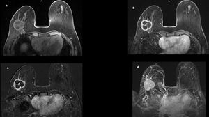
Fig. 1: Invasive ductal carcinoma of the right breast
A 55-year-old female with a lesion in the right breast.
(a) Axial 3D T1-weighted fat-sat images reveal an irregular lobulated and heterogeneous mass. (b) Axial 3D T1-weighted fat-sat images with gadolinium show a centrally low signal mass, consistent with the necrotic part of the tumour with peripheral rim enhancement. (c) Subtracted image. (d) Maximum intensity projection image (MIP)
References: Department of radiology, IASO Hospital
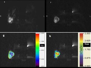
Fig. 2: Invasive ductal carcinoma of the right breast
(e) b0 image of DWI sequence. (f) b1200 image shows the lesion as a high signal mass.
(g) corresponding Kapp map and (h) Dapp map
References: Department of radiology, IASO Hospital
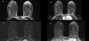
Fig. 3: Fibroadenoma of the left breast
A 45-year-old female with a lesion in the left breast. (a) Axial 3D T1-weighted fat-sat images reveal an isointense well circumscribed ovoid mass. (b) Axial 3D T1-weighted fat-sat images with gadolinium show a circumscribed mass with homogeneous enhancement. (c) Subtracted image. (d) Maximum intensity projection image (MIP)
References: Department of radiology, IASO Hospital
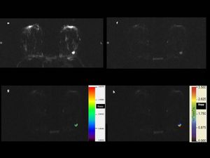
Fig. 4: Fibroadenoma of the left breast
(e) b0 image of DWI sequence. (f) b1200 image shows the lesion as a high signal mass.
(g) corresponding Kapp map and (h) Dapp map
References: Department of radiology, IASO Hospital
Image Analysis
All breast MR images were interpreted on a workstation by an experienced radiologist.
The operator was blinded to the final diagnosis.
The first post contrast subtracted images were used primarily to identify and localize suspicious lesions according to their initial enhancement.
The MIP images were used to clarify whether there was any significant enhancement.
The radiologist reviewed the images using an in-house processing routine that allows the placement of 3D volumes-of-interest (VOIs) incorporating voxels across multiple slices.
The axial high b-value,
the Kapp and Dapp maps were viewed concurrently in a single session for each case.
The median Kapp and Dapp values were then calculated from the histogram from the whole-lesion.
MRI offers a high sensitivity for breast cancer and has traditionally been used as a second-line imaging method to solve diagnostic problems in patients with equivocal findings on mammography or ultrasound and recently as a screening method to improve prognosis of the women at increased risk of breast cancer [1-13].
However,
the full diagnostic protocol is time and cost consuming.
The early arterial phase after contrast injection is best suited to visualize the enhancement of the invasive breast cancers as well as the ductal carcinoma in situ (DCIS).
The remaining dynamic phases and additional pulse sequences of current MRI protocols have traditionally been acquired not to detect but rather to characterize enhancing structures.
Based on this rationale the abbreviated MRI protocol has been suggested in order to reduce the time and the cost of the MRI [14].
However,
it is sometimes impossible to distinguish enhancing benign tumors from malignant carcinomas unambiguously based on the contrast enhancement alone and it is been reported that the specificity of the dynamic contrast enhanced (DCE) MRΙ is suboptimal,
around 75% [15,16].
DKI model provides a novel contrast mechanism in MRI and has a high sensitivity in the detection of changes in the local biologic environment.
Diffusion-weighted MRI (DWI) is based on the gaussian diffusion model assuming that water molecules diffuse uniformly in a certain direction.
In real tissue with complex cellular structures,
the water molecules within an imaging voxel (typically 2 Χ 2 Χ 2 mm) diffuse through an environment that is highly heterogeneous in any direction.
DKI model includes the kurtosis term to capture this deviation from the gaussian distribution.
Changes in DKI parameters may reflect physiologic and morphologic alterations associated with breast tumor tissues; thus,
DKI may add valuable indications of microstructural changes to conventional diffusion techniques for the characterization of the breast tumors [17,18].
Statistical Analysis
Histological analysis of the breast lesions was performed and further comparative analysis of the results was done to investigate the accuracy of the method.
The Kapp and Dapp median values between benign and malignant lesions were compared by the Mann-Whitney U test.
Thelevel of significance set at 0.05.
Receiver operating characteristics (ROC) analysis was used to determine the most effective cutoff values for the differentiation between benign and malignant pathologies.
All analyses were performed by the use of MedCalc 15.0 software.
